- The paper introduces a dual-path diffusion framework that jointly estimates global homography and local deformations for precise cross-modal retinal image alignment.
- It employs transformer-based and CNN-based score networks with iterative refinement and input-adaptive guidance to overcome disparities in field-of-view and image quality.
- ADM demonstrates superior performance over state-of-the-art methods by achieving higher mAUC and robust convergence on challenging SFI-UWFI datasets.
Active Diffusion Matching: A Score-based Iterative Approach for Cross-Modal Retinal Image Alignment
Introduction
Aligning standard fundus images (SFIs) and ultra-widefield fundus images (UWFIs) is a critical but challenging task in ophthalmic image analysis. These modalities display substantial disparities in field-of-view (FOV), scale, color, and local retinal features, making cross-modal registration highly nontrivial. Prior methods either only address SFI-SFI registration or rely on hand-crafted keypoints and affine transformations, which are inadequate given the SFI-UWFI domain gap. The "Active Diffusion Matching: Score-based Iterative Alignment of Cross-Modal Retinal Images" paper (ADM) (2604.10084) introduces a novel, diffusion-based framework that iteratively estimates both global transformations and local deformations, thereby enabling robust, fully automated SFI-UWFI alignment.
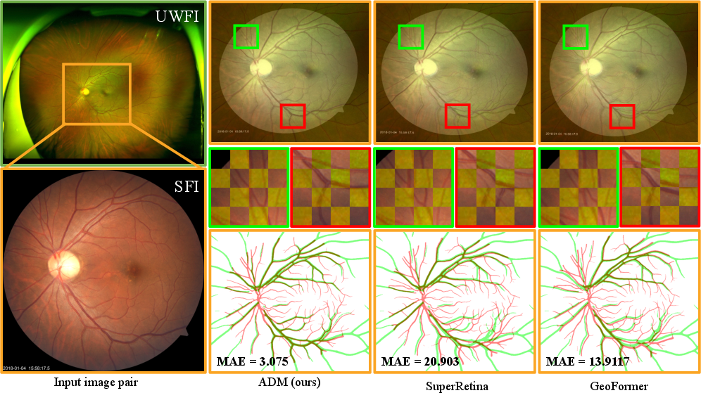
Figure 1: SFI-UWFI alignment via ADM, with FOV differences and comparative local alignment results versus SuperRetina and GeoFormer.
Methodology
Dual-path Score-based Diffusion Model
ADM builds upon recent advances in score-based generative models and Langevin Markov chains, simultaneously leveraging two independent yet interdependent diffusion models. These models jointly estimate:
- Global Homography (H): Handled by a transformer-based score network, modeling the dominant projective transformation between SFI and UWFI domains.
- Local Displacement Field (v): Captured via a CNN-based (U-net) score estimator, modeling local deformation beyond the global homography.
Each model is trained with a conditional denoising score-matching loss, enabling progressive refinement through a reverse diffusion process. Critically, inference is performed by iteratively updating estimates of Ht and vt in a coupled manner, utilizing a customized guidance scheme based on appearance consistency.
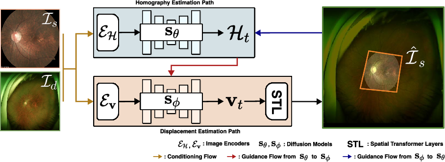
Figure 2: ADM overview—source (SFI) and dest (UWFI) images are aligned via dual score networks for homography (sθ) and local field (sϕ), conditioned through learned encoders and combined via spatial transformer layers.
Additional salient aspects include:
- Input-adaptive guidance: At each inference step, ADM modulates the score prediction for homography (sθ) by incorporating gradients of pixel-level appearance loss, enhancing adaptability to challenging image pairs.
- Iterative refinement: The aligned output is recursively re-used as input, further minimizing residual misalignment.
- Dynamic scheduling: Losses and guidance terms are weighted adaptively across the diffusion trajectory to encourage stability and convergence.
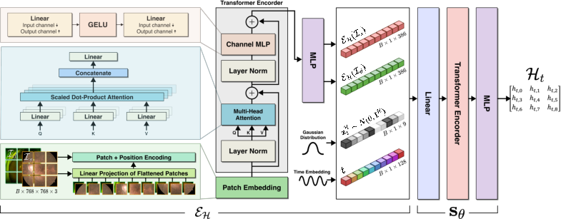
Figure 3: Homography estimation path architecture—image features via EH and transformer-based predictor sθ.
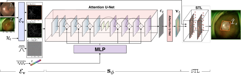
Figure 4: Displacement field estimation path—vessel-enhanced features via Ev and U-net-based v0 yield local deformation maps.
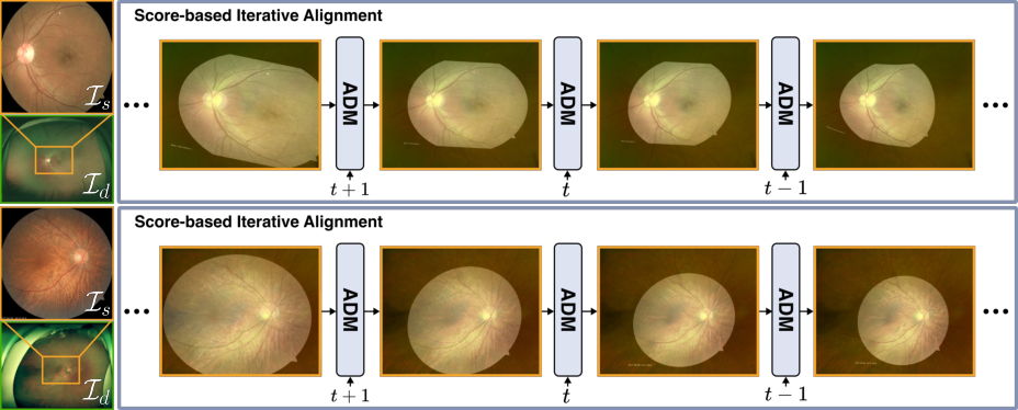
Figure 5: Score-based iterative alignment—progressive prediction and composition of global and local alignment transformations.
Experimental Results
Datasets
ADM's efficacy is validated on two benchmarks:
- KBSMC (private): 3744 SFI-UWFI pairs exhibiting strong real-world variability in scale and FOV.
- FIRE (public): 134 SFI-SFI pairs, standard benchmark for retinal image registration.
Key metrics are maximum alignment error (MAE), median error (MEE), and mean Area Under the Curve (mAUC) for acceptable registration.
Quantitative Results
ADM demonstrates superior performance on the challenging KBSMC dataset:
| Method |
Acceptable Rate (%) |
Inaccurate (%) |
mAUC |
| GeoFormer |
36.10 |
63.90 |
24.1 |
| MCNet |
32.89 |
67.11 |
20.9 |
| ADM |
41.98 |
58.02 |
29.3 |
On FIRE, ADM matches or slightly surpasses top-performing SFI-SFI baselines with highest mAUC reported.
Qualitative Results
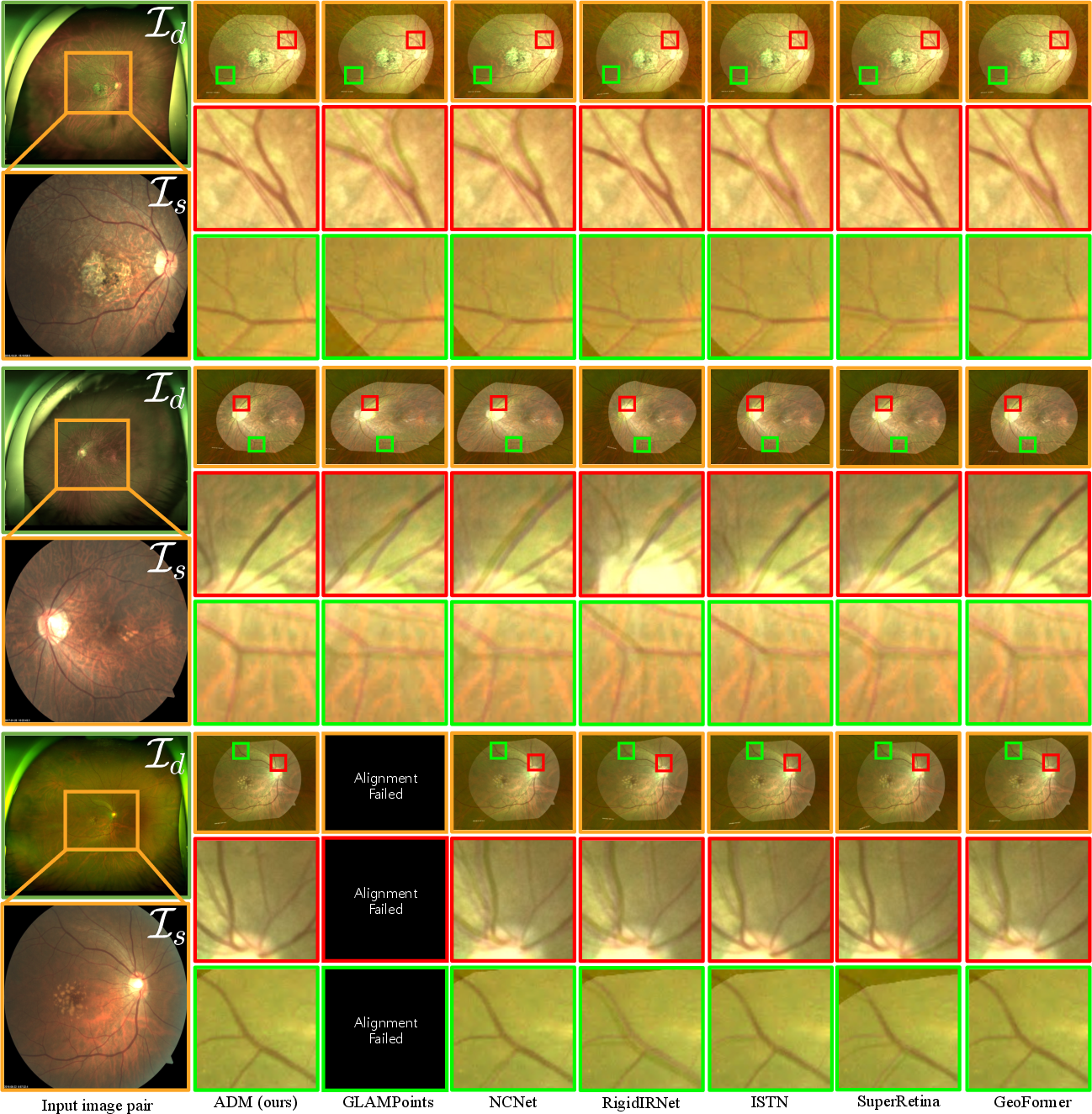
Figure 6: Direct homography estimation qualitative comparison—ADM achieves superior alignment compared to GLAMPoints, NCNet, RigidIRNet, ISTN, SuperRetina, and GeoFormer on SFI-UWFI pairs.
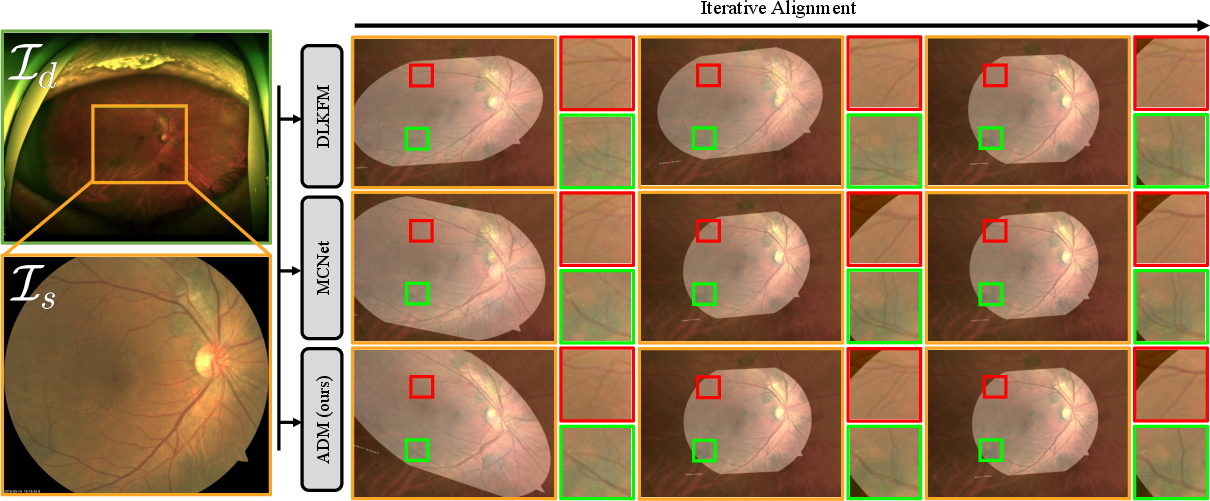
Figure 7: Iterative homography refinement—ADM attains more accurate and faster convergence than DLKFM and MCNet.
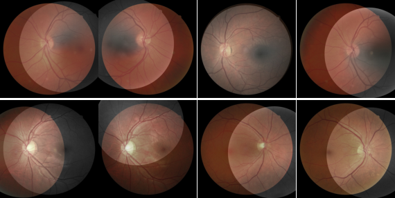
Figure 8: ADM aligns SFI-SFI image pairs in the FIRE dataset robustly, with accurate overlay of anatomical structures.
Ablation and Analysis
ADM's architectural and algorithmic choices are systematically ablated:
- Guided sampling and iteration: Removing adaptive guidance or iterative updates significantly degrades mAUC and alignment accuracy.
- Network design: The transformer-for-global, CNN-for-local split provides superior results compared to alternatives.
- Dynamic loss weighting: Adaptive schedules for key loss terms stabilize global transformation estimation.
ADM also demonstrates robustness to Gaussian noise, blur, and low illumination, with only moderate performance drops.
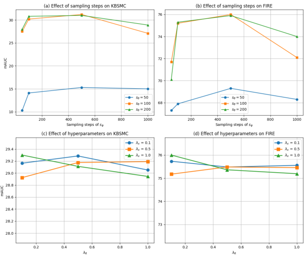
Figure 9: Ablative evaluation on ADM iterations and hyperparameters—performance saturates with increased sampling steps for global estimator; local estimator is sensitive to oversmoothing.
Limitations and Failure Cases
Despite its strengths, ADM's extended inference time (≈47 s per pair) is substantially slower than some baselines due to iterative Langevin steps. Furthermore, in highly degraded scenarios with low vessel visibility, the local deformation path (v1) may fail, analogous to keypoint-based approaches under low SNR.
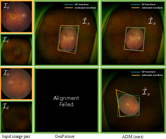
Figure 10: Failure cases—ADM and GeoFormer both fail under severe blur and low illumination where anatomical structure extraction is unreliable.
Theoretical and Practical Implications
The integration of dual-path diffusion modeling provides a theoretically grounded framework for disentangling global and local transformations in cross-modal registration. ADM's architecture is extensible: any medical imaging task requiring multimodal registration with significant domain gaps stands to benefit. The stochastic iterative formulation enables adaptation to previously unseen test cases beyond what is achievable with feedforward architectures.
Practically, aligning SFI-UWFI pairs enables the construction of large datasets for image enhancement and super-resolution, potentially elevating UWFI to the gold standard for retinal assessment with minimal diagnostic compromise. Robust cross-modal registration is also fundamental for multimodal fusion and longitudinal studies in ophthalmology.
Future Directions
Potential avenues for extending ADM’s capabilities include:
- Acceleration: Adoption of fast or one-step diffusion sampling with knowledge distillation [song2023consistency, salimans2022progressive].
- Enhanced robustness: Integration of adversarial training or hybrid fusion with keypoint-based cues; dynamic preprocessing for quality-aware vessel extraction.
- Generalization: Application to other anatomical domains (e.g., brain MRI-to-CT registration) and 3D multimodal data.
Conclusion
ADM introduces a diffusion-based, dual-path framework for cross-modal retinal image alignment, outperforming conventional and state-of-the-art deep learning baselines on SFI-UWFI alignment tasks. Its joint treatment of global and local transformation estimation sets a new standard for robust, adaptable medical image registration, with broad implications for downstream clinical applications and cross-modal data fusion (2604.10084).