- The paper demonstrates a novel pipeline for pulmonary vascular extraction using multi-scale Hessian filter fusion and TEASAR skeletonization that bypasses the need for annotated voxel data.
- It fuses Frangi and Sato filters to improve continuity of both distal microvasculature and central arteries, validated by morphometric metrics such as fractal dimension and Murray’s exponent.
- The resulting vascular graph exhibits enhanced topology with increased nodes and bifurcations, offering practical insights for quantitative pulmonary imaging and computational modeling.
Spatial Graph Representation and Morphometric Analysis of Pulmonary Vasculature from CT via Multi-Scale Filter Fusion and TEASAR Skeletonization
Methodological Overview
The paper presents a deterministic, training-free pipeline for the spatial graph-based reconstruction and morphometric analysis of the pulmonary vascular tree from non-contrast chest CT data. The approach circumvents the need for voxel-wise annotated datasets typical of deep learning-driven vessel segmentation, leveraging multi-scale Hessian-based vesselness filtering fused from Frangi and Sato responses, advanced morphological preprocessing, and TEASAR-based skeletonization. The input consists of isotropically-resampled chest CT scans acquired on a SPECT/CT platform with compromised spatial resolution and elevated noise, thus simulating a challenging clinical context.
The lung segmentation protocol implements HU thresholding, morphological operations to sever airway continuity, and Chan-Vese active contour refinement on reduced resolution slices, capped by exclusion of voxels proximate to the lung boundary to suppress pleural artifacts.
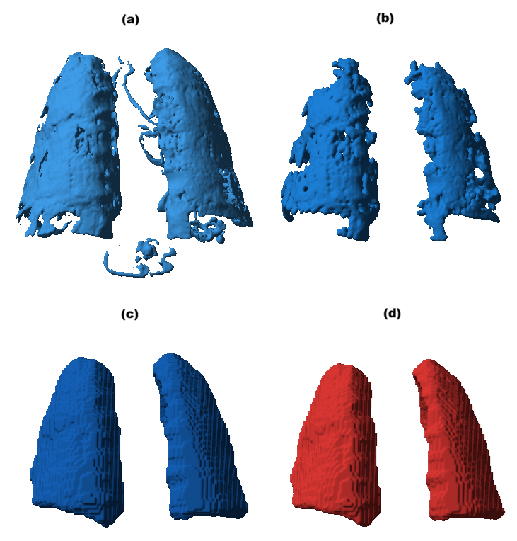
Figure 1: Sequential stages of the lung segmentation pipeline, progressing from HU thresholding to airway disconnection, Chan-Vese refinement, and pleural exclusion.
Subsequently, the vesselness enhancement module applies Frangi and Sato filters across twelve spatial scales ($1$-$8$ mm), exploiting their complementary sensitivity profiles: Frangi excels in distal, narrow branches but is discontinuous on large vessels; Sato resolves central arteries continuously but is insensitive to peripheral microvasculature. Fused vesselness maps employ a weighted maximum operator, slightly favoring the Sato response, followed by percentile-based normalization and thresholding.
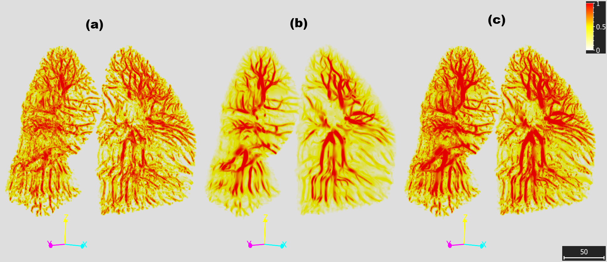
Figure 2: Comparative visualization of Frangi, Sato, and fused vesselness responses highlighting the complementary strengths of each and demonstrating the fusion's capacity to simultaneously capture distal and central branches.
A close-up analysis of vessel continuity elucidates the fusion's utility in bridging gaps in Frangi that Sato fills, thus restoring uninterrupted vessel topology.
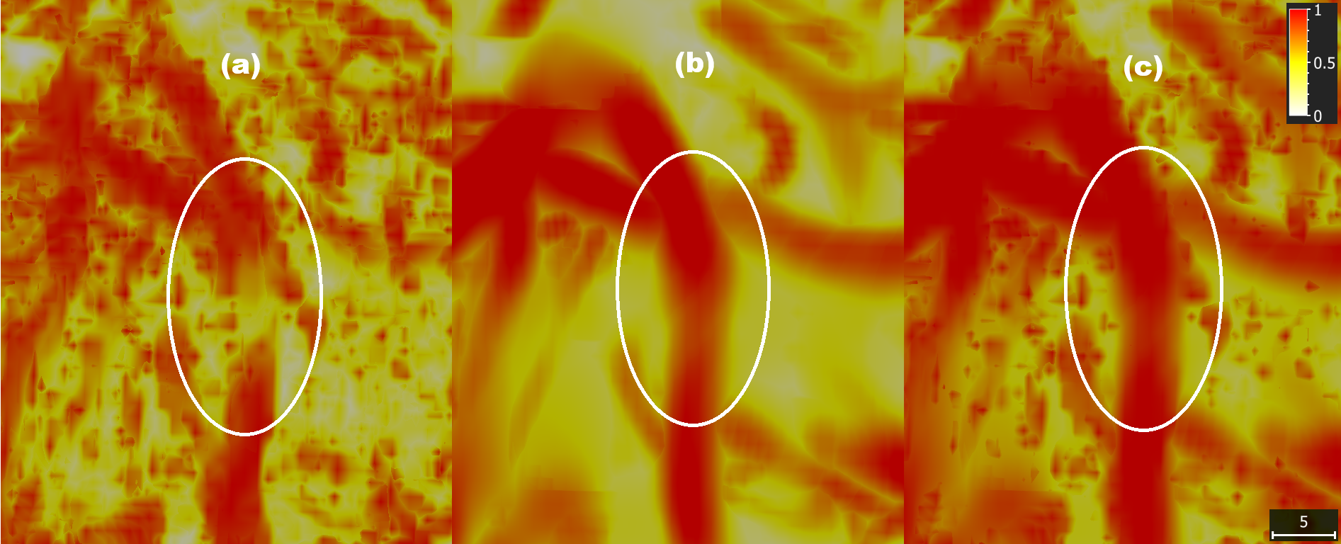
Figure 3: A medium pulmonary vessel as seen under the three vesselness filters, showing continuity restoration in the fusion.
Skeletonization proceeds via Kimimaro's TEASAR implementation, tracing centerlines and extracting per-vertex radii from EDT, followed by graph construction, pruning, and acyclicity validation, ensuring anatomical plausibility. Graph edges encode physical branch paths and radii, and are classified topologically.
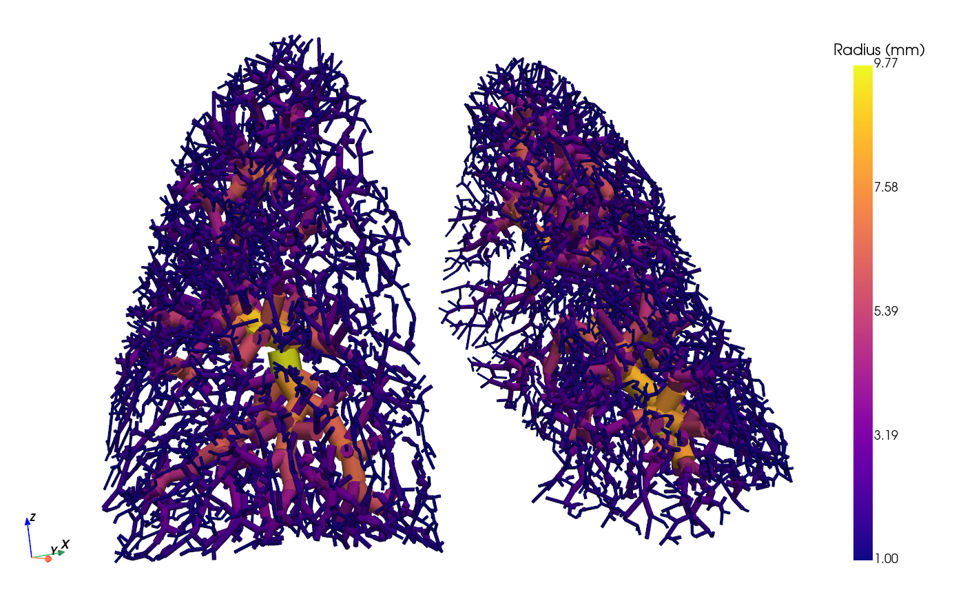
Figure 4: The final spatial vascular graph—nodes marking junctions, paths, and endpoints.
Morphometric Characterization
Morphometric analysis employs established descriptors to validate the anatomical and geometric plausibility of reconstructed vascular graphs: 3D box-counting for fractal dimension, Murray's law exponent α estimation via nonlinear minimization, Strahler ordering, Horton ratios, branch tortuosity, and bifurcation asymmetry.
Fractal dimension (volumetric, Dfvol) quantifies the vascular tree's space-filling capacity, evaluated on skeletons inflated with per-node radii. Murray's law validation involves log-log regression of parent versus summed daughter radii at bifurcations, targeting α within physiological bounds. Strahler ordering and Horton ratios assess self-similarity and branching patterns, while tortuosity and asymmetry characterize geometric features at the segment and bifurcation levels.
Fusion critically increases the reconstructed graph's topological reach (nodes, bifurcations), raises Dfvol, and recovers both large-vessel continuity and distal microvasculature as compared to pure Frangi or Sato runs.
(Figure 5: Figure ID not provided; See Table 1 in paper for numerical comparison.)
(Figure 6: Figure ID not provided; Fractal and Murray's law fits; refer to detailed results.)
Numerical Results and Contradictory Claims
The weighted fusion of Frangi and Sato filters produces a vascular graph with 12,929 nodes and 4,520 bifurcations, 53% and 28% greater than Frangi and Sato, respectively. Dfvol (2.26±0.05) falls strictly within clinical ranges reported for human pulmonary vasculature. Murray's exponent (α=1.81±0.03) aligns with empirical measurements in human pulmonary arteries, differing substantially from the classical optimum (α=3), with Frangi showing higher $8$0 due to superior distal vessel capture. Horton ratios and Strahler order maxima reflect expected truncations from limited CT resolution, with bifurcation ratios close to anatomical literature norms but inflated length ratios due to fragmentary peripheral segments.
The claim that the fusion outperforms both individual filters in terms of morphometric reconstruction fidelity is substantiated by quantitative topology and space-filling complexity metrics. Notably, the reported asymmetry ratios are higher than reference values, a consequence of missing distal branches, which may bias bifurcation statistics toward symmetry.
Practical and Theoretical Implications
The pipeline demonstrates that deterministic, training-free vessel segmentation and spatial graph modeling from clinical CT can yield morphometrically plausible pulmonary vascular trees, supporting downstream applications such as quantitative morphometry, computational lung modeling, and ventilation-perfusion assessment without reliance on large annotated datasets or deep learning infrastructure.
Practically, this methodology enables rapid, transparent vascular tree extraction in resource-limited or heterogeneous clinical environments where training data is scarce, scanner protocols vary, and reproducibility is essential. The explainable nature and tunable parameters facilitate adaptation to variable CT quality and anatomical variation.
Theoretically, the deviation in Murray's exponent and bifurcation asymmetry from classical optimality, compounded by truncation of deepest Strahler orders, signals the influence of CT spatial resolution on physiologically informed morphometric analysis. Further, the fusion strategy reveals a tractable path to multi-scale vessel segmentation, accommodating the pulmonary tree's profound diameter heterogeneity.
Future developments may involve:
- Extension to high-resolution and contrast-enhanced CT,
- Clinical cohort validation,
- Integration with deep learning pipelines for hybrid approaches,
- Automated parameter adaptation,
- In silico modeling of hemodynamic properties from spatial graphs.
Conclusion
This research introduces a robust, transparent pipeline for pulmonary vascular segmentation and spatial graph-based morphometric analysis from clinical CT, achieving plausible anatomical and geometric characterizations consistent with empirical physiological benchmarks despite poor input resolution. Weighted fusion of multi-scale Frangi and Sato filters is critical for joint recovery of central and peripheral vasculature, underpinning accurate spatial graph topology and complexity. Limitations include single-patient scope and absence of ground truth, but the method is immediately applicable for quantitative pulmonary imaging and has broad translational potential in computational modeling, morphometric biomarker discovery, and explainable image analysis.