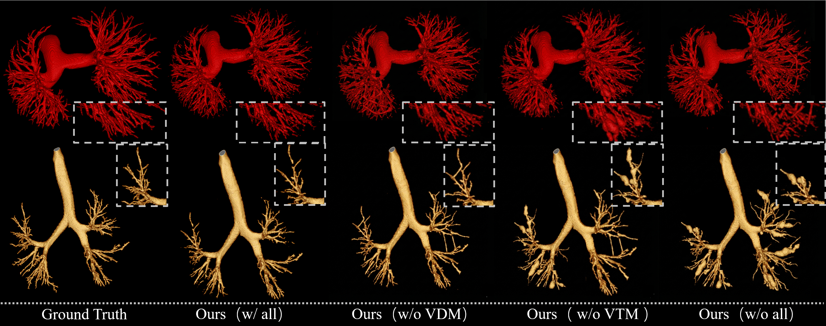
- The paper presents a novel framework that jointly predicts vessel masks, distance maps, and thickness maps to enforce geometric consistency and boundary precision.
- It adapts a frozen SAM ViT encoder with a lightweight 2.5D adapter to efficiently capture volumetric context while maintaining resource efficiency.
- Experimental results on Parse2022 and AIIB23 benchmarks show significant improvements in Dice and clDice scores, demonstrating enhanced segmentation performance.
MorVess: Morphology-Aware Pulmonary Vessel Segmentation Network
Motivation and Background
Accurate segmentation of pulmonary vessels in CT remains an unsolved challenge due to sparse and tortuous vasculature, multiscale branching, and low signal-to-noise ratios especially in distal microvasculature. Conventional segmentation models, primarily optimizing binary masks, inadequately capture topological integrity and geometric consistency of vascular trees. They frequently exhibit discontinuities, lose small or peripheral branches, and fail to enforce physiologically plausible vessel diameter gradients under voxel-wise supervision. Post hoc topology-preserving loss terms such as clDice partially mitigate these issues, but lack continuous geometric guidance during learning, limiting their impact.
MorVess introduces an integrated framework coupling explicit geometric priors with efficient adaptation of large foundation models to volumetric medical imaging. Joint prediction of vessel masks, distance maps (VDM), and thickness maps (VTM) instantiates a regime where semantic and geometric supervision are tightly coupled, reinforcing vascular connectivity and boundary precision. The design achieves high-fidelity vessel reconstruction via adaptation of a frozen SAM ViT encoder to 3D contexts, fused with multi-level geometric and semantic cues.
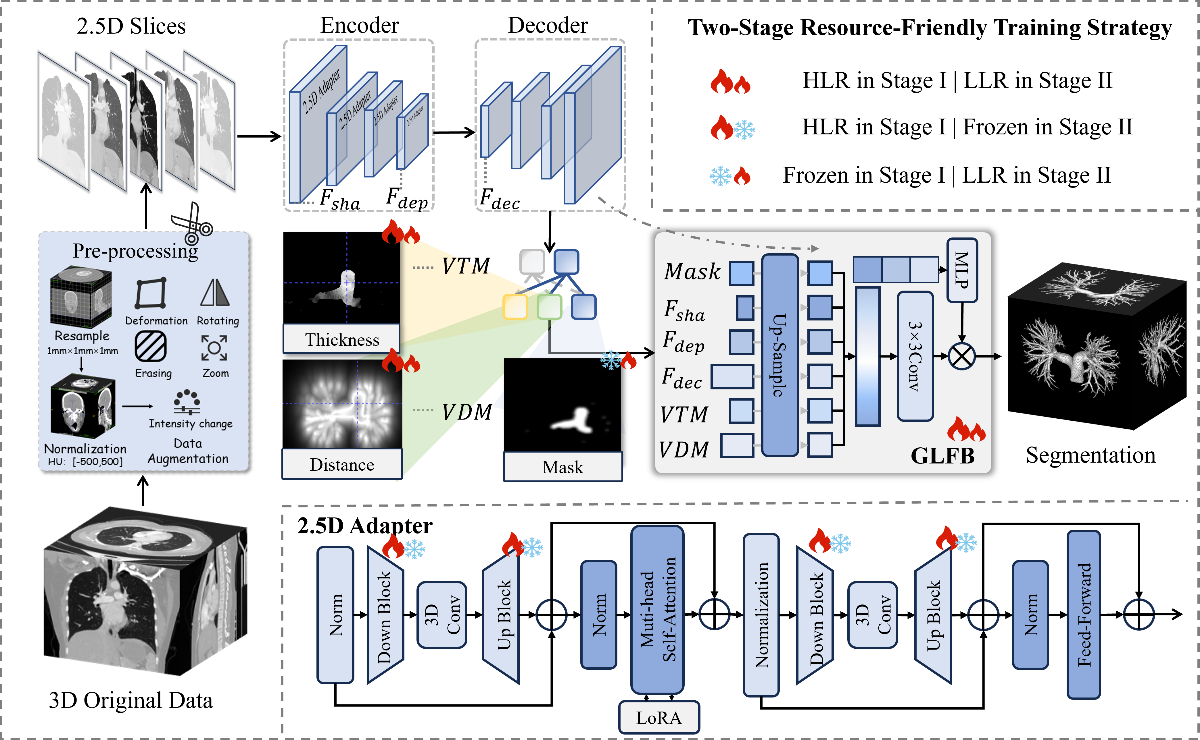
Figure 1: Schematic of MorVess with 2.5D adapter, multi-head decoder, and global-local fusion integrating multi-scale semantics and geometric fields.
Geometric Priors: Construction and Role
The geometric prior module generates two differentiable fields:
- Vessel Distance Map (VDM): Encodes decaying potential from vessel boundaries, mitigating boundary ambiguity and enabling sub-voxel boundary localization. Boundary voxels are treated as high-energy layers, and exponentially decaying distance fields regularize segmentation with superior boundary adherence.
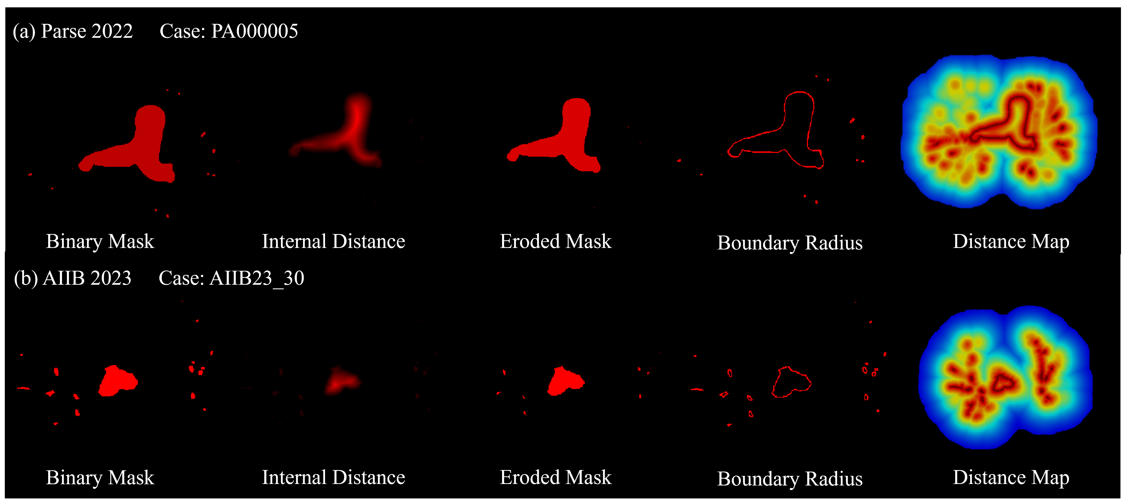
Figure 2: Generation of VDM via morphological erosion and exponential distance decay from vessel masks.
- Vessel Thickness Map (VTM): Governs diameter consistency by propagating maximal inscribed sphere radii from skeletonized centerlines. This enforces smooth diameter transitions and suppresses non-physical oscillations, critical for topological continuity in distal vasculature.
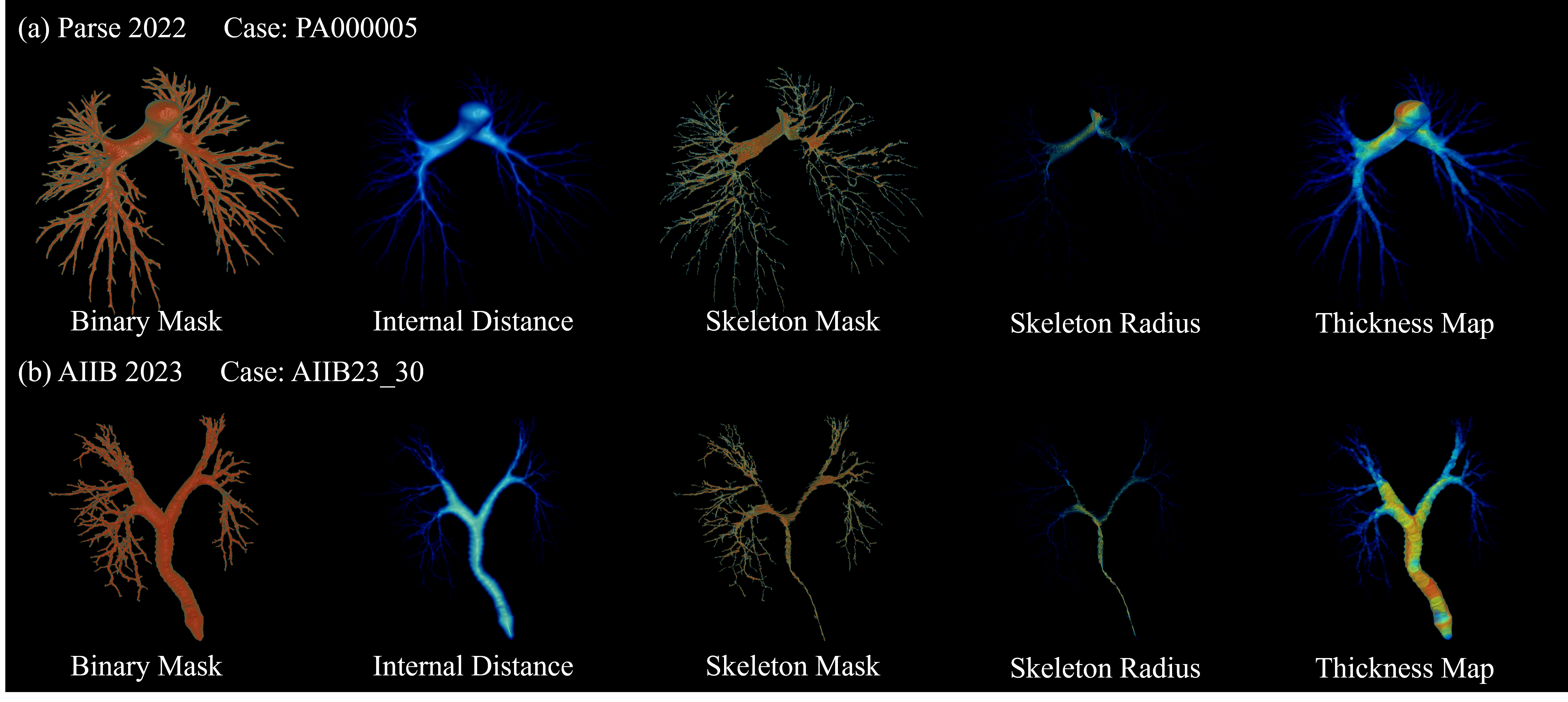
Figure 3: VTM creation by skeleton extraction and medial-axis propagation, assigning thickness to each vessel voxel.
These priors are generated for each ground-truth mask and predicted jointly with segmentation outputs, establishing a composite loss function combining CE, Dice, clDice, VDM, and VTM constraints.
Architecture: SAM Adaptation and Fusion
MorVess leverages the SAM ViT encoder as a foundation, adapted to volumetric medical domains via a lightweight 2.5D adapter. This module models Z-axis dependencies between slices using efficient 3D convolutions, enabling minimal parameter overhead while bridging 2D/3D context. Only the Adapter and decoder branches are trained, with the ViT backbone frozen, resulting in an efficient architecture with negligible resource inflation.
A multi-head decoder generates semantic masks, VDM, and VTM in parallel. A Global-Local Fusion Block (GLFB) consolidates shallow and deep encoder features, decoder outputs, and geometric priors, enabling adaptive channel-wise weighting via vessel tokens and a hypernetwork. This mechanism corrects local errors and enhances recognition of fine branches and boundaries.
Experimental Evaluation
MorVess was validated on Parse2022 and AIIB23 benchmarks, both characterized by complex and pathologically distorted vascular networks. Standard preprocessing includes voxel intensity normalization, overlapping slice stacking, and ground-truth prior map generation. The network is trained in two stages: macro-feature adaptation of the Adapter and GLFB, then fine-tuning of geometric branches.
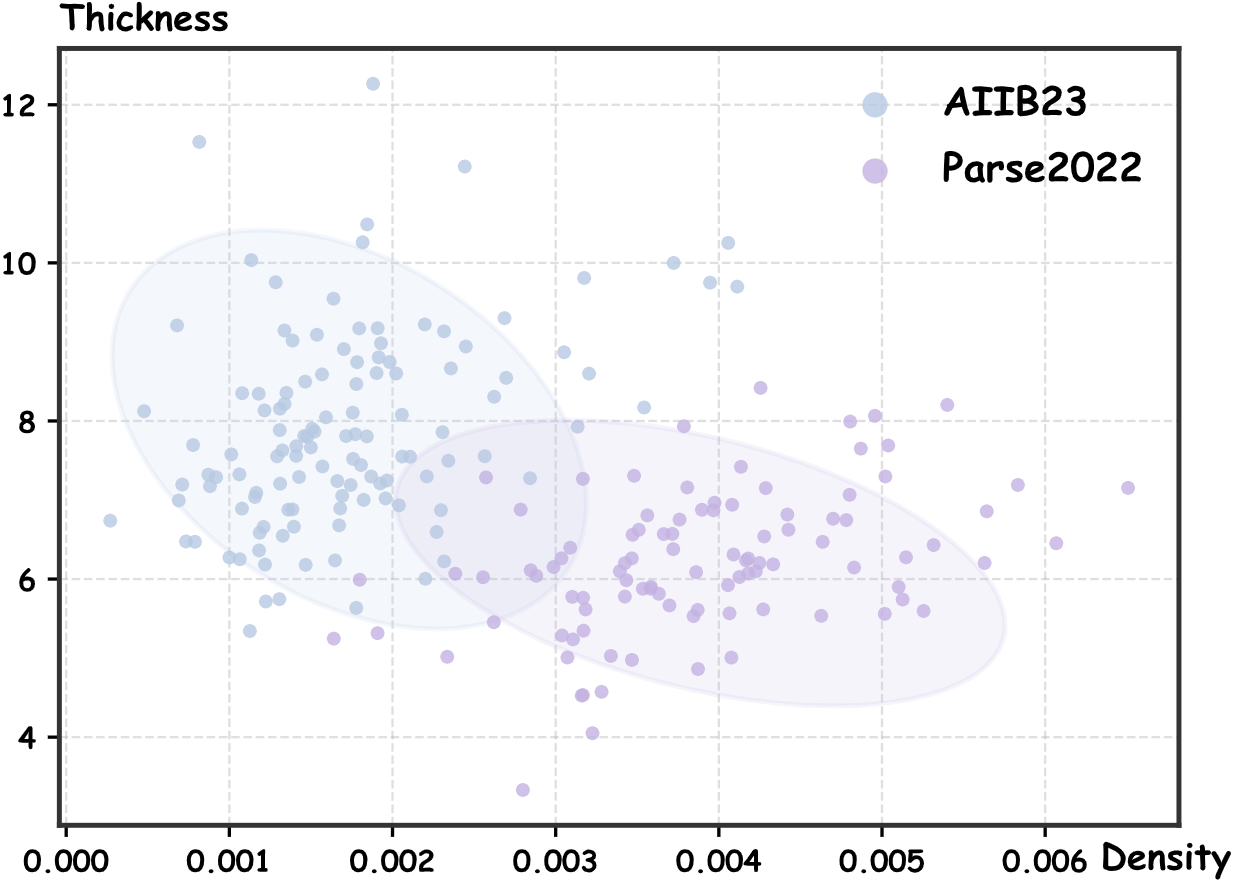
Figure 4: Feature space scatter demonstrating vascular density and thickness distribution for Parse2022 and AIIB23.
MorVess achieved superior scores across Dice, clDice, HD95, branch/length ratios (DBR/DLR), and apparent missing rate (AMR), showing statistically significant gains over all baselines. For example, Dice improved by almost 10% over Swin-UNETR (86.84% vs. 76.85%) and clDice outperformed COMMA (83.22% vs. 80.10%) on Parse2022. On AIIB23, it reached 94.31% Dice and 89.34% clDice, outperforming all competitors under difficult conditions.
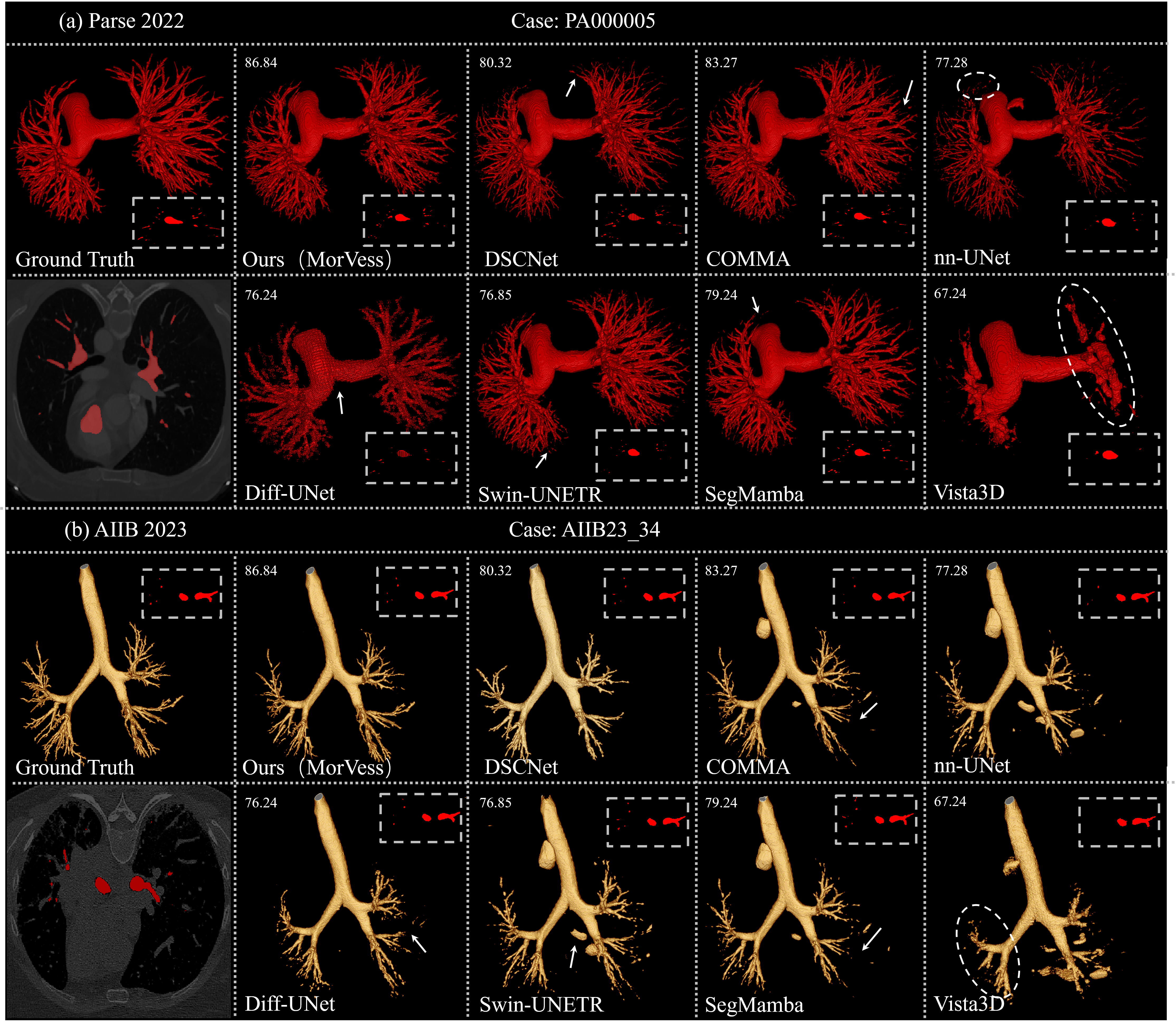
Figure 5: 3D qualitative comparison with SOTA on Parse2022 and AIIB2023; MorVess maintains continuity in challenging regions.
Ablation Studies
Ablations confirm the synergistic role of geometric priors:
Key component removal (2.5D Adapter, GLFB, SAM pretraining) demonstrated their necessity: the Adapter and GLFB yield complementary gains, and SAM pretraining further boosts performance.
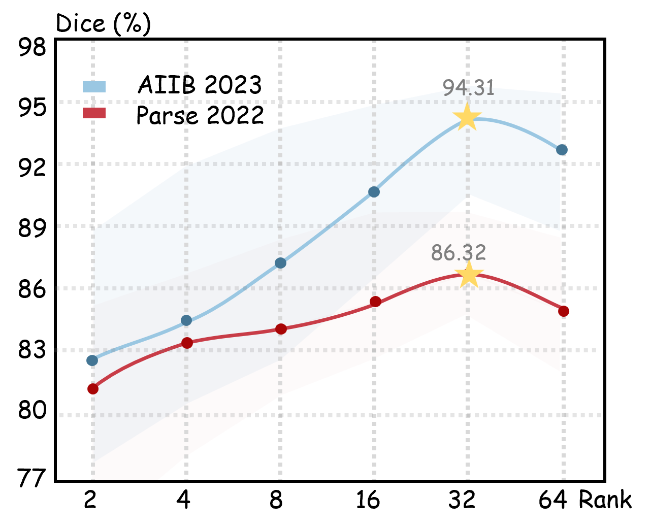
Figure 7: Dice scores saturate at FaCT rank 32, balancing representational capacity and parameter efficiency.
MorVess is highly resource-efficient: total trainable parameters are 1.0 M (versus 32–64 M for nnU-Net/Diff-UNet), 42 GMACs per slice stack, and 4.2 GB memory peak (vs. 18–32 GB). These results confirm high performance is attainable with minimal computational overhead.
Domain Generalization and Geometric Consistency
Cross-dataset experiments show MorVess generalizes across arterial/venous and peripheral domains without fine-tuning, retaining global topology and achieving only minor drops in Dice/clDice. VTM-based supervision captures geometric invariants, supporting robustness to anatomical variance.
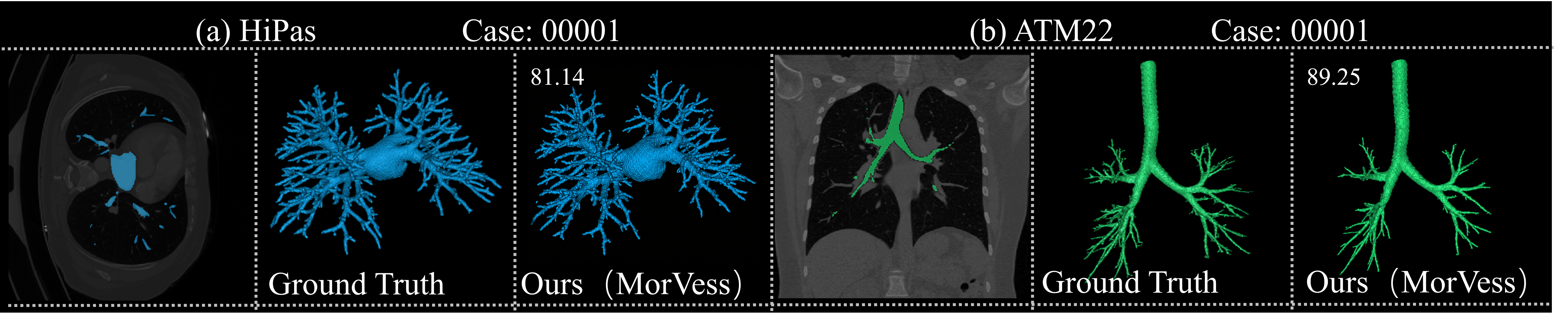
Figure 8: Qualitative cross-domain results on HiPas and ATM2022; continuity is retained in out-of-distribution settings.
Geometric analyses using VMTK evaluate total vessel volume (TVV), diameter distribution, and small-vessel volume fraction. MorVess exhibits lowest TVV error and highest Pearson correlation for diameter histograms, while small-vessel fidelity is substantially improved, supporting accurate modeling for clinical hemodynamics and remodeling research.
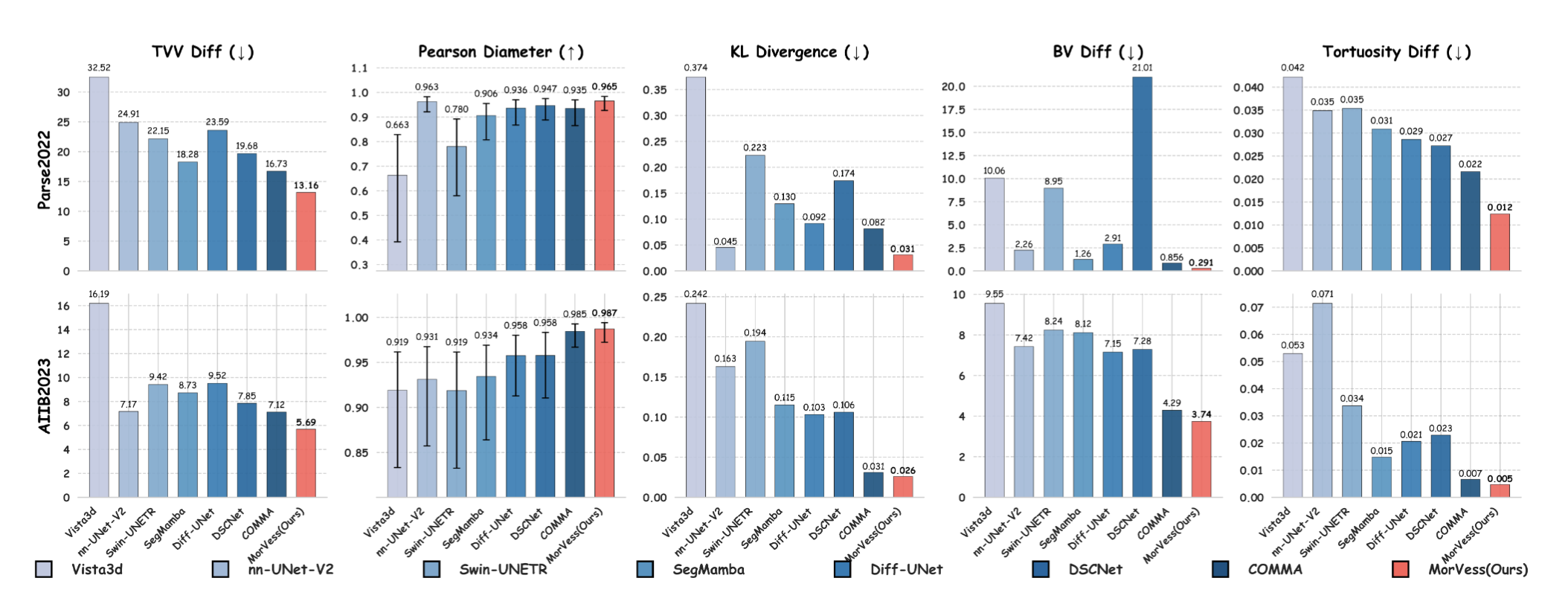
Figure 9: Geometric consistency analysis via centerline extraction; MorVess minimizes TVV error and preserves diameter gradients and distal branch volume.
Limitations and Future Directions
MorVess’s 2.5D adaptation limits Z-axis receptive field to ±2 slices, restricting capture of long-range spatial dependencies and robustness to anisotropic CT. Future work will explore attention-based mechanisms with extended axial context and cross-site validation.
Conclusion
MorVess establishes an explicit geometric prior segmentation paradigm, coupling differentiable anatomical fields with pre-trained visual foundation models for anatomically faithful, resource-efficient pulmonary vessel segmentation. Joint modeling of masks, VDM, and VTM enforces topological integrity, boundary precision, and diameter consistency, supporting high-fidelity parsing across domains. These contributions have potential to accelerate clinically reliable vascular quantification and facilitate subsequent functional modeling and disease diagnostics in pulmonary imaging.