- The paper demonstrates that context-aware evaluation and multi-tumor generalization are crucial for robust mitosis detection, evidenced by a drop in mean precision from 0.805 in hotspots to 0.400 in challenging areas.
- The study employs a spectrum of methodologies including single-stage object detectors with ensembling and parameter-efficient adaptations, underscoring architectural diversity in overcoming domain bias.
- The findings stress that clinical applications must validate detection models on random and challenging whole-slide regions rather than solely on curated hotspots.
Context-Aware Multi-Tumor Generalization: The MIDOG 2025 Challenge
Introduction
Automating mitotic figure (MF) detection in histopathology images remains a fundamental but unsolved problem for computational pathology due to substantial biological and contextual variability. The MIDOG 2025 challenge, as detailed in "Mitosis Detection in the Wild: Multi-Tumor and Context-Aware Generalization in the MIDOG 2025 Challenge" (2606.07368), constitutes the most ambitious evaluation to date, acknowledging that reliable clinical deployment of MF detection requires robustness across tumor types and spatial contexts far beyond previous benchmarks. Unique to MIDOG 2025 is the extension of evaluation from expert-selected hotspots to random and challenging whole-slide regions across an expanded set of human and veterinary tumor domains.
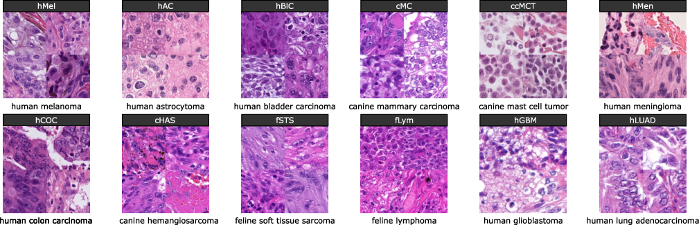
Figure 1: Domains of the test set of the MIDOG 2025 challenge, showing four example hotspot-region samples per domain across 12 tumor types representing marked biological diversity.
Challenge Structure and Dataset Properties
MIDOG 2025 consisted of two tracks: (1) object detection of mitotic figures in region-of-interest (ROI) images; and (2) binary classification of MFs as normal or atypical. The test set included 365 ROIs sampled from 122 whole-slide images representing 12 tumor types—spanning human, canine, and feline pathology—and digitized on diverse scanner models. Crucially, sampling strategies encompassed classical hotspots, uniformly random regions reflective of whole-tissue diversity, and intentionally challenging areas saturated with MF imposters.
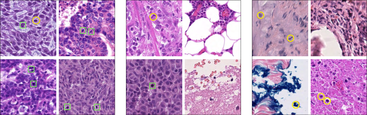
Figure 2: Region types for Track 1, illustrating hotspot (left), random (middle), and challenging (right) ROIs; imposters yielding reference model false detections are circled in yellow.
The ROI-level MF distribution confirms dramatic context heterogeneity, with hotspot regions, random regions, and challenging regions presenting sharply contrasting MF prevalence and difficulty.
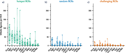
Figure 3: Violin plots of MF distributions for hotspot, random, and challenging ROIs, showing substantially reduced MF counts and increased detection difficulty in challenging and random areas.
For atypical mitosis classification in Track 2, the test set reflected severe class imbalance, also varying by tumor type.
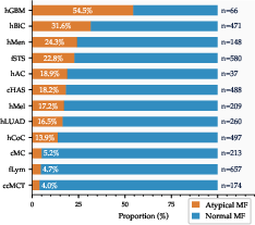
Figure 4: Class distribution by tumor type in the Track 2 test set, revealing atypical MF underrepresentation in most domains.
Methodological Overview
A broad methodological spectrum characterized the challenge submissions. In Track 1, a predominant trend was the usage of single-stage object detectors (YOLOv5/8/10/11/12, RTMDet, DETR), with a minority leveraging segmentation-based (nnUNet, VM-UNET) or two-stage architectures. Secondary classifiers, test-time augmentation (TTA), and ensembling were variably employed. Training data strategies primarily revolved around the MIDOG++ dataset, with some leveraging auxiliary datasets providing greater context or domain diversity.
In Track 2, the leading approaches integrated parameter-efficient adaptation (LoRA) of vision foundation models (DINOv3, Virchow, UNI, HIBOU) as well as ConvNeXt, EfficientNet/ViT, and a range of ensembling paradigms. Fine-tuning on the MIDOG++ and newly released atypical mitosis datasets enabled strong performance across tumor and scanner domains.
Main Results and Performance Analysis
Track 1: MF Detection
Top-performing algorithms reached F1 scores up to 0.740. However, there was an explicit collapse in model precision outside curated hotspot regions: mean precision fell from 0.805 (hotspot) to 0.400 (challenging areas), with the false positive rate increasing by 208% in difficult contexts. Recall values remained relatively stable, implicating inadequate model specificity as the prime failure mode in challenging tissue scenarios.
Score analyses demonstrated that hotspot performance is not predictive of generalization, with Pearson r = 0.36 (CI: -0.206, +0.750; p=0.201) between hotspot and challenging ROI performance. Performance also varied significantly by tumor type, e.g., F1 ranging from 0.444 (human glioblastoma) to 0.808 (canine hemangiosarcoma):
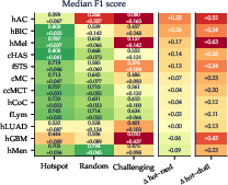
Figure 5: MF detection F1 score by tumor type and region, highlighting pronounced domain-blind spots.
Track 2: Atypical Mitosis Classification
Challenge-winning classifiers achieved balanced accuracy of 0.908 (Balezo et al.), with ROC-AUC and BA uniformly exceeding prior benchmarks. Class imbalance and tumor domain effects remained acute, with feline lymphoma domains presenting the greatest difficulties due to low atypical MF recall.

Figure 6: Balanced Accuracy and ROC AUC distribution by tumor type, reflecting both cross-domain strengths and residual weaknesses.
Ablation and Efficiency Insights
A systematic ablation study showed that ensembling delivered a mean absolute gain of 1.5 percentage points in F1 for detection and 1.3 for balanced accuracy in classification, especially for challenging areas, indicating that model uncertainty and fractional specialization are more valuable as the distribution diverges from training data. In contrast, TTA yielded negligible or even negative impact, questioning its continued use despite community prevalence.
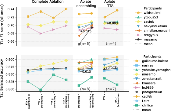
Figure 7: Ablation study quantifying performance deltas for TTA and ensembling in both challenge tracks.
Inference speed and VRAM usage demonstrated broad variance. Notably, high-performing submissions were not necessarily the most computationally intensive or slowest, indicating efficient architectures can remain performant.
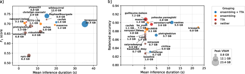
Figure 8: Inference time versus main metric for both tracks, with bubble size indicating VRAM; no monotonic relation between speed/resource use and performance is seen.
Practical and Theoretical Implications
The MIDOG 2025 results clearly demonstrate that current deep learning models exhibit a context bias, with strong performance in curated hotspots failing to generalize to realistic or adverse tissue contexts. This confirms a critical limitation in clinical WSI deployment: models validated on hotspot-only datasets cannot be assumed reliable for broad tissue analysis.
Practically, this argues for mandatory evaluation—including random and challenging regions—prior to any clinical adoption of MF detection tools. The demonstrated superiority of ensembling in domain-divergent regions and the diminishing return of TTA advocate for efficient model curation and community-wide reporting of ablation studies. The dominance of foundation model adaptation (LoRA) in atypical classification establishes a pragmatic path for rapid cross-domain generalization, but also reinforces data-centric curation as a bottleneck—especially concerning region-level context and rare tumor types.
Theoretically, these findings motivate algorithmic innovations that transcend context bias and exploit spatial tissue priors. Domain adaptation, unsupervised context modeling, or uncertainty-aware segmentation-classification cascades may provide incremental robustness. The challenge outcome suggests foundation model scaling is not a substitute for explicit spatial and context-aware learning.
Future Directions
Future research should prioritize:
- Systematic dataset curation embedding context and rare tumor class diversity for both detection and classification
- Architectural research targeting explicit spatial context encoding and imputed region bias
- Investigations of uncertainty quantification for deployment gating and region triage
- Expansion of evaluation frameworks to prioritize generalization, nuanced error analyses, and robustness under domain shift
Direct applicability of vision foundation models (with LoRA or fine-tuning) and high-performing YOLO-based detectors should be contextualized by their fragility in challenging and rare-domain scenarios. Further, prospectively defined benchmarks with full-slide inference and clinical workflow integration will be essential to move computational pathology beyond algorithmic contests toward translational impact.
Conclusion
MIDOG 2025 provides the most granular and comprehensive assessment of mitosis detection and atypical mitosis classification methods available, revealing the persistent domain and context sensitivity of current state-of-the-art models. Clinical deployment should not be contemplated without robust validation in representative, non-hotspot, and challenging contexts. The challenge's open-access datasets and well-defined benchmarks now constitute a critical resource to drive foundational advances in context-aware pathology AI.