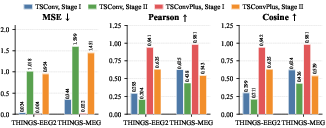
- The paper introduces a dual-stage framework that uses a spatio-temporal VAE and Q-Former to bridge visual and M/EEG signal domains.
- It demonstrates significant performance improvements, achieving subject-specific Pearson correlations of 0.635 for EEG and 0.543 for MEG.
- The method offers actionable insights for individualized BCI and neuroprosthetic applications while effectively addressing neural variability.
Authoritative Summary of “ViBE: Visual-to-M/EEG Brain Encoding via Spatio-Temporal VAE and Distribution-Aligned Projection”
Introduction and Motivation
Visual brain encoding is fundamental for understanding how external visual stimuli are processed into neural activity, contributing both to systems neuroscience and the development of neural prosthetic devices. Recent progress in deep learning has facilitated new models for fMRI-based encoding, but with limited practicality for BCI, due to cost and temporal constraints. Electro- and magnetoencephalography (M/EEG), by contrast, enable rapid, noninvasive neural readout, yet high-fidelity visual-to-M/EEG encoding remains challenging due to the complexity of the spatio-temporal neural dynamics and the significant distributional gap between visual and neural representations.
Existing methods predominantly rely on deterministic mappings (Bao et al., 4 Mar 2025, Mai et al., 14 Aug 2025), which cannot capture the one-to-many mapping structure and neural variability intrinsic to M/EEG responses. Direct alignment between high-level visual embeddings (e.g., from CLIP) and neural latent spaces is further confounded by severe feature scale misalignment, resulting in suboptimal reconstructions and poor cross-modal correspondence.
Methodology
ViBE addresses these limitations with a two-stage cross-modal generative framework, emphasizing hierarchical spatio-temporal modeling and probabilistic alignment. The architecture is structured as follows.
Stage I: The TSC-VAE (Spatio-Temporal Separated Convolutional Variational Autoencoder) is designed to reconstruct M/EEG signals by combining temporal convolutions (1,kt) and spatial convolutions (ks,1) with ks<C, where C is the number of spatial channels. This design (TSConvPlus) preserves hierarchical spatial information, in contrast to methods that collapse spatial dimensions. The VAE objective combines standard MSE reconstruction and a KL bottleneck.
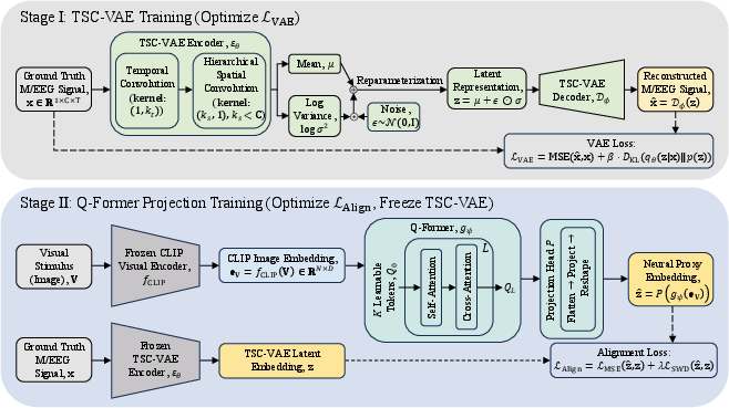
Figure 1: Overview of the two-stage ViBE training procedure, showing the TSC-VAE (Stage I) and cross-modal Q-Former alignment (Stage II).
Stage II: Q-Former receives CLIP image embeddings and uses learnable query tokens with cross-attention to extract visual-semantic features, which are projected into the TSC-VAE latent space (“neural proxy embeddings”). A combined MSE and sliced Wasserstein distance (SWD) loss aligns neural proxy and VAE latent embeddings, aligning both pointwise and distributional structure.
Inference Pipeline
At inference, a novel image is processed by the frozen CLIP encoder, Q-Former, and the TSC-VAE decoder to generate subject-specific or subject-generalized M/EEG signal predictions.

Figure 2: End-to-end ViBE inference: image → CLIP embedding → Q-Former → TSC-VAE Decoder → predicted M/EEG signal.
Experimental Results
ViBE is systematically evaluated on THINGS-EEG2 and THINGS-MEG, with extensive baselines, including MindSimulator (Bao et al., 4 Mar 2025), SynBrain (Mai et al., 14 Aug 2025), and previous CLIP-based methods (2509.00787). Three key metrics are compared: MSE (lower is better), Pearson correlation, and cosine similarity.
Strong claims in the results:
Modality Gap and Cross-Modal Alignment
Statistical analysis of embedding distributions reveals a substantial input-output scale gap: CLIP image features are ∼100(ks,1)0 smaller in variance than M/EEG latent embeddings. The Q-Former brings the proxy embedding scale within (ks,1)12.5(ks,1)2 of the neural distribution, dramatically improving alignment and thus cross-modal mapping.
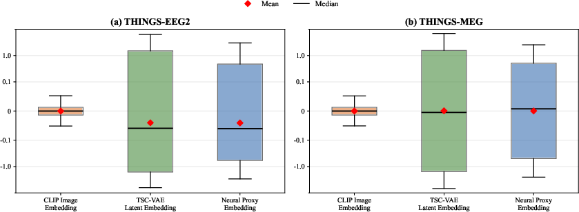
Figure 4: Distributional comparison of CLIP, TSC-VAE, and neural proxy embeddings, highlighting effective scale bridging by Q-Former.
Loss ablations confirm that including the SWD term, even at the cost of slight MSE increases, robustly enhances matched structure as measured by Pearson and cosine scores.
Neuroanatomical Analysis
Brain region ablations substantiate the critical role of occipital and temporal regions (“visual pathway”) in visual signal representation. Selective occlusion of these regions yields the largest performance decrements, in line with known neuroscience principles.
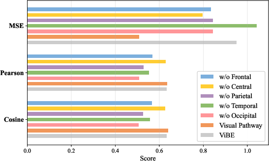
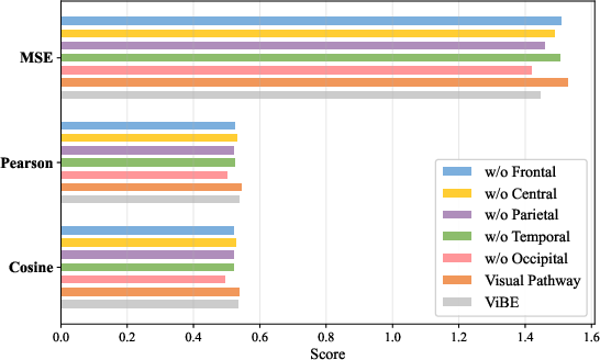
Figure 5: Brain region ablation results: removing occipital/temporal locations degrades signal prediction, affirming their representational importance.
Implications and Future Work
ViBE’s subject-specific and multi-subject protocols set a new reference for high-fidelity, image-conditioned brain encoding in temporally resolved modalities. The explicit separation and bridging of modality scales enables distributional matching that is essential for robust cross-modal generative modeling. Practically, these advances support more accurate, individualized BCI and neuroprosthetics pipelines capable of leveraging high-throughput neural signals, while theoretically, the work motivates deeper alignment modeling (scale, shape, and context). Outstanding questions include scaling to larger, more diverse populations, investigating task generalization, and integrating adaptive normalization layers to further reduce scale mismatch.
Conclusion
ViBE presents a rigorously validated and technically well-motivated approach to visual-to-M/EEG encoding, featuring (i) spatio-temporally aware variational autoencoding, (ii) cross-modal scale bridging via Q-Former, and (iii) joint alignment losses. The method demonstrates strong improvements in signal reconstruction quality across challenging datasets and advances the methodological frontier for brain encoding and prosthetic applications.