WarpPINN: Cine-MR image registration with physics-informed neural networks
Published 22 Nov 2022 in eess.IV, cs.CV, and cs.LG | (2211.12549v1)
Abstract: Heart failure is typically diagnosed with a global function assessment, such as ejection fraction. However, these metrics have low discriminate power, failing to distinguish different types of this disease. Quantifying local deformations in the form of cardiac strain can provide helpful information, but it remains a challenge. In this work, we introduce WarpPINN, a physics-informed neural network to perform image registration to obtain local metrics of the heart deformation. We apply this method to cine magnetic resonance images to estimate the motion during the cardiac cycle. We inform our neural network of near-incompressibility of cardiac tissue by penalizing the jacobian of the deformation field. The loss function has two components: an intensity-based similarity term between the reference and the warped template images, and a regularizer that represents the hyperelastic behavior of the tissue. The architecture of the neural network allows us to easily compute the strain via automatic differentiation to assess cardiac activity. We use Fourier feature mappings to overcome the spectral bias of neural networks, allowing us to capture discontinuities in the strain field. We test our algorithm on a synthetic example and on a cine-MRI benchmark of 15 healthy volunteers. We outperform current methodologies both landmark tracking and strain estimation. We expect that WarpPINN will enable more precise diagnostics of heart failure based on local deformation information. Source code is available at https://github.com/fsahli/WarpPINN.
The paper introduces WarpPINN, a physics-informed neural network that accurately registers cine-MRI and computes myocardial strain using hyperelasticity constraints.
It employs Fourier feature mapping to overcome spectral bias, resulting in precise displacement field reconstruction and sub-3mm median landmark errors.
Quantitative results demonstrate enhanced strain estimation and deformation field preservation compared to traditional methods and CNN-based approaches.
Physics-Informed Cardiac MRI Registration: The WarpPINN Framework
Introduction and Problem Context
Quantification of local cardiac deformations, particularly myocardial strain, is increasingly recognized as critical for advanced assessment of heart function, especially since standard global metrics such as ejection fraction often fail to discriminate phenotypes of heart failure. Cine steady-state free precession (SSFP) MRI is the clinical standard for cardiac imaging but lacks direct encodings of tissue motion, making noninvasive quantification of regional strain challenging. The paper presents a physics-informed neural network (PINN) approach, termed WarpPINN, to infer the cardiac deformation field from cine-MRI for robust image registration and strain mapping, leveraging the near-incompressibility of myocardial tissue.
Model Architecture and Physics-Informed Loss
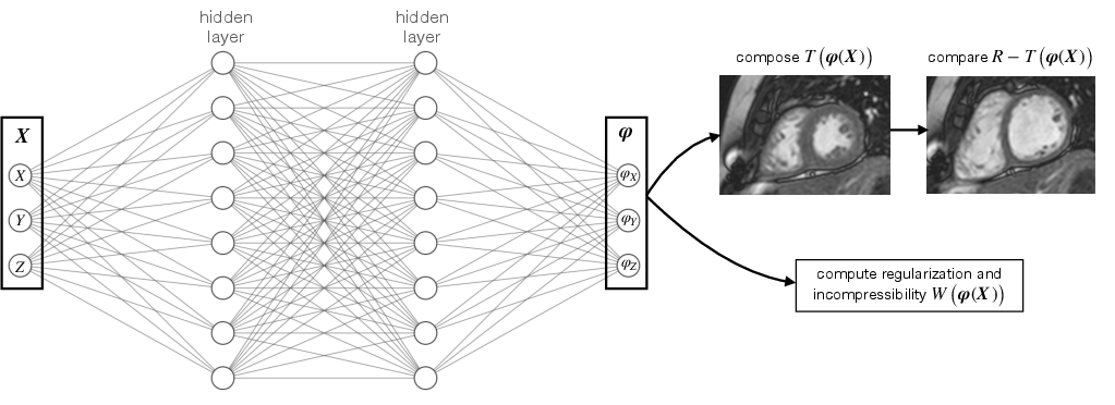
WarpPINN represents the spatial deformation field φ:Ω→Rn (with n=2,3) via a fully connected neural network that regresses displacements u(X;θ), so that φ(X;θ)=X+u(X;θ). The network is trained to minimize a loss combining an intensity-based similarity term and a physically motivated regularizer:
L(θ)=∥R−T∘φ∥pp+μR(φ)
Here, R is the reference (end-diastolic) image, T is a template image, and the regularizer R derives from hyperelasticity theory (specifically, a neo-Hookean law) to penalize deviation from incompressibility in identified myocardial regions. The network enables computation of the full strain tensor via automatic differentiation.
Figure 1: A neural network predicts the displacement field φ as a function of the position in the image X, optimizing image registration under near-incompressibility constraints.
Addressing the Spectral Bias: Fourier Feature Mapping
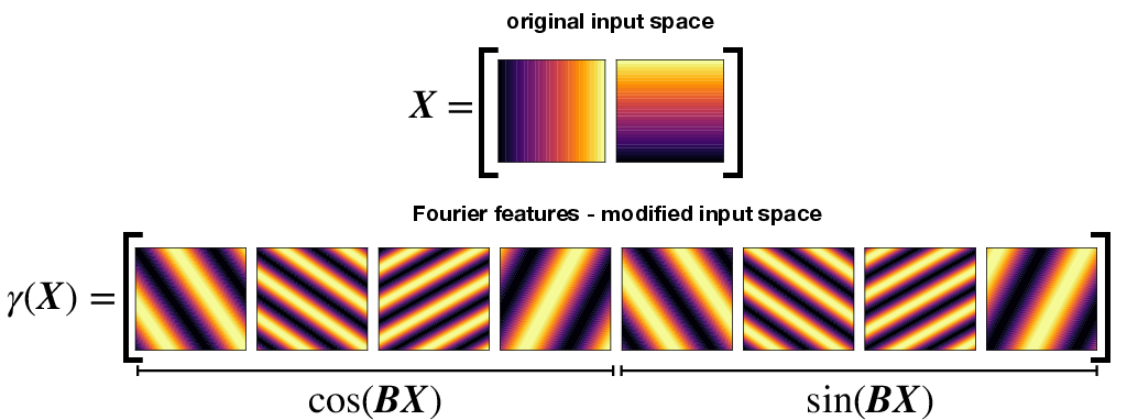
A salient challenge for PINN-based regression of deformation fields is the spectral bias of standard feedforward networks, manifesting as poor approximation of high-frequency or discontinuous real displacement fields, e.g., at tissue interfaces. WarpPINN addresses this by augmenting the input layer with randomized Fourier feature mappings:
γ(X)=[cos(BX),sin(BX)]
where B is a fixed Gaussian random matrix. This modification enables the network to efficiently express high-frequency spatial features and discontinuities.
Figure 2: Fourier features help overcome the spectral bias of neural networks by introducing modified input space with higher frequencies.
Synthetic 2D Validation: Registration and Strain Recovery
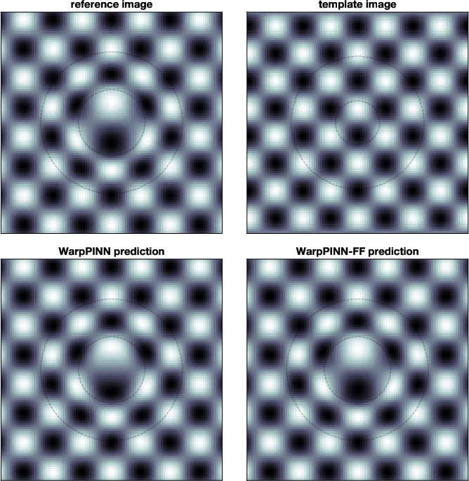
A controlled synthetic experiment is designed wherein a 2D image with a ring-shaped incompressible region undergoes a known deformation, producing reference and template images. Training the PINN—with and without Fourier features—demonstrates that Fourier augmentation dramatically improves the accuracy of both the displacement field and, crucially, its (potentially discontinuous) derivatives relevant for strain computation.
Figure 3: Reference and template images (top), and the warped predictions using WarpPINN and WarpPINN-FF (bottom) illustrating improved reconstruction with Fourier features.
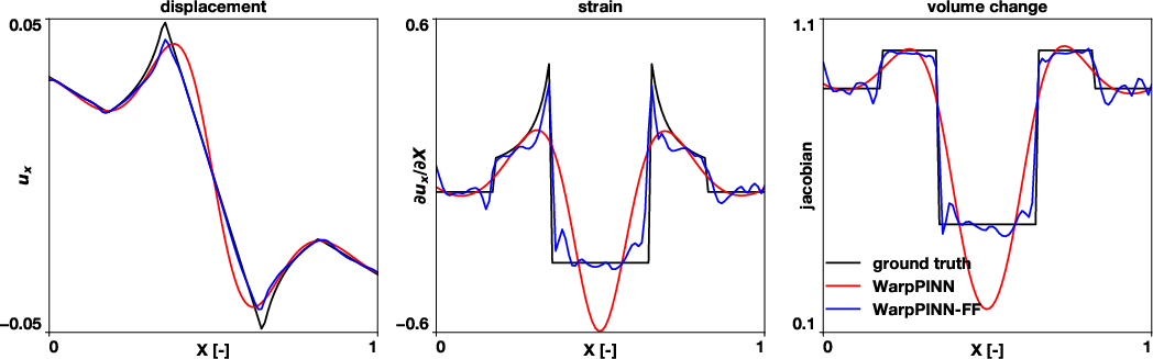
Figure 4: Profiles at y=0.505 of ux, ∂ux/∂X, and Jacobian, confirming that WarpPINN-FF better captures discontinuities in derivatives.
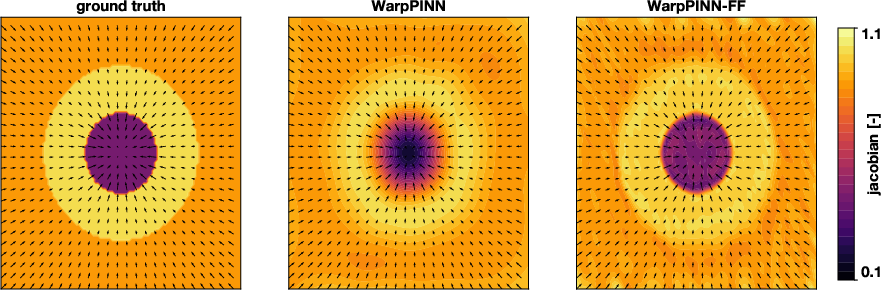
Figure 5: Ground truth jacobian versus PINN and PINN-FF reconstructions; PINN-FF captures spatial sharpness while the standard PINN excessively smooths.
Benchmarking on Cine-MRI Data
WarpPINN is further evaluated on the Cardiac Atlas Project's multimodal motion tracking dataset, comprising cine-MRI for 15 healthy volunteers with manual landmark annotations and mesh-based region segmentations.
Key elements of the practical implementation:
The 3D (spatial) + time (t) deformation field is parameterized by the neural network, with [X,t] as input.
Training uses mini-batch sampling over both data points and regularization collocation points, exploiting mesh segmentations to selectively apply incompressibility enforcement in the myocardium and not in the background.
Template images correspond to different frames along the cardiac cycle.
Deformation fields are used to advect annotated landmarks and predict their motion.
Numerical Performance and Strain Mapping Results
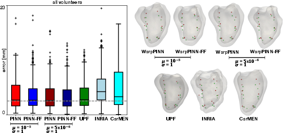
Landmark Tracking: WarpPINN and its Fourier-augmented variant achieved landmark position errors with a median as low as 2.91 mm, outperforming or equalling established conventional (UPF, INRIA) and CNN-based (CarMEN) approaches for given regularization hyperparameters.
Figure 6: Left—box plots for landmark tracking errors, with WarpPINN-FF (μ=5×10⁻⁶) exhibiting the lowest median; Right—deformed mesh with predicted and ground truth landmarks.
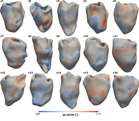
Deformation Field Quality: Visualization of jacobian determinants at end-systole over all volunteers demonstrates preservation of near-incompressibility (J≈1 across myocardium) as prescribed by the physics-informed regularization, with PINN-FF showing better handling of larger deformations but increased oscillations in some edge cases.
Figure 7: Jacobian (determinant of deformation gradient) at end-systole, as predicted by WarpPINN for all volunteers.
Figure 8: Jacobian fields predicted by WarpPINN-FF at end-systole, revealing larger permissible deformations compared to standard PINN.
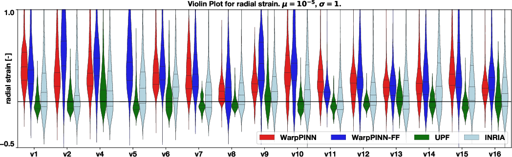
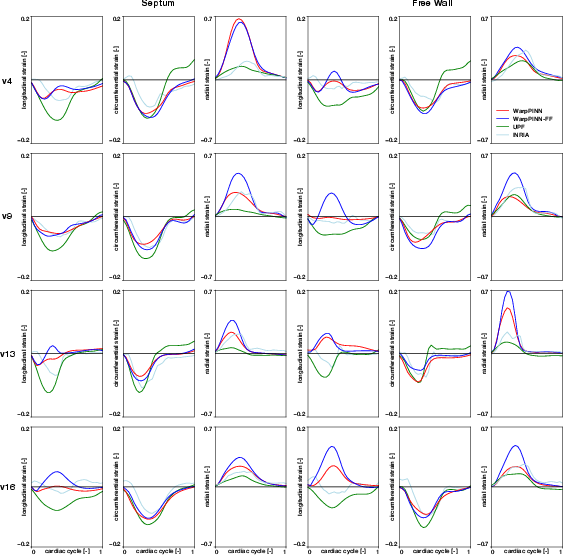
Strain Quantification: Computed regional radial strain distributions show higher physiological dynamic range versus UPF and INRIA for radial strains (up to 45%), with temporal smoothness beneficial for tracking cardiac phases and minimizing drift.
Figure 9: Violin plots for radial strain at end-systole in all volunteers, with WarpPINN-FF producing a wider, more physiological distribution.
Figure 10: Strain curves for four volunteers, showing high peaks in radial strain and temporal smoothness for both PINN and PINN-FF versus UPF and INRIA.
Discussion and Comparative Analysis
Why Physics-Informed Learning?
Imposing biophysical constraints via a PINN uniquely enables continuous, differentiable deformation fields that respect the underlying mechanics—here, near-incompressibility of myocardium under loading. The resulting fields are not only physically plausible but generalize across sparse and noisy regimes, outperforming classical intensity-based regularization in biomechanical realism and clinical utility of strain fields.
Benefits of Fourier Features
Fourier feature mappings facilitate the learning of highly non-smooth deformation fields, crucial for capturing real anatomical discontinuities, at the cost of potential overfitting for large frequency hyperparameters. Moderate tuning is required to balance flexibility with stability.
Regularization Trade-offs
The regularization parameter μ governs a trade-off between registration fidelity (lower μ) and physically plausible, less distorted deformation (higher μ). Smaller μ values yield lower landmark errors but risk excessive volume changes and unphysical strains; higher μ values may under-fit true tissue motion, particularly in outlier frames or volunteers.
Practical Considerations
Resource Requirements: Training WarpPINN for a full cardiac sequence on 3D+t images with a single case requires substantial computational resources (NVIDIA QUADRO RTX 8000, 70–140 min per case).
Transferability: The model is currently case-specific, with ongoing work needed on transfer learning and “one-shot” encoding of deformation across cases.
Comparison with Other Methods: CNNs (CarMEN) enable batch processing across cases but often lack explicit physical constraints, leading to less reliable strain estimation.
Implications and Future Directions
WarpPINN establishes a pipeline for direct, physics-regularized estimation of clinically relevant deformation fields and cardiac strain metrics from widely available cine-MRI, potentially enabling earlier and more specific detection of regional cardiac dysfunction. The methodology is immediately extensible to other organ systems and image modalities, contingent on knowledge of relevant tissue mechanics.
On a theoretical level, this work motivates integration of invertibility guarantees (e.g., through invertible neural networks) to ensure true diffeomorphic mappings, improved loss functions for better outlier resistance, and automated hyperparameter tuning (e.g., Bayesian optimization). Incorporating uncertainty quantification and transfer learning may further bridge the gap toward robust population-wide and personalized deployment.
Conclusion
WarpPINN presents a robust, interpretable, and extensible framework for cardiac image registration and strain mapping, leveraging physics-informed neural networks and spectral input augmentation. The method achieves sub-3mm median landmark errors and physiologically plausible strain ranges superior to traditional methods, with potential for significant impact on quantitative cardiac diagnostics, clinical research, and broader biophysical modeling tasks.
“Emergent Mind helps me see which AI papers have caught fire online.”
Philip
Creator, AI Explained on YouTube
Sign up for free to explore the frontiers of research
Discover trending papers, chat with arXiv, and track the latest research shaping the future of science and technology.Discover trending papers, chat with arXiv, and more.