- The paper introduces covariance-guided graph deformation to reconstruct volumetric cardiac meshes with high boundary accuracy.
- It employs a joint 3D CNN-GNN architecture with template-driven warping to ensure topology consistency and robust simulation fidelity.
- Quantitative results show lower Chamfer distances and zero inverted elements, underscoring its effectiveness for in-silico trials.
Covariance-Guided Cardiac Volumetric Mesh Reconstruction: The HeartVolMesh Framework
Motivation and Problem Statement
High-fidelity, topology-consistent tetrahedral meshes are critical for cardiac digital twin construction, enabling robust FEM/CFD simulations for in-silico trials. Prevailing segmentation-then-meshing pipelines introduce blurred anatomical boundaries and stochastic cross-case vertex connectivity, limiting mesh correspondence, population analysis, and standardized learning/simulation workflows. Prior deformation-based image-to-mesh models (e.g., Voxel2Mesh, MeshDeformNet, HeartDeformNet) enforce deterministic Euclidean supervision and produce surface-only outputs that require post hoc volumetric tetrahedralization, further breaking volumetric correspondence and mesh regularity.
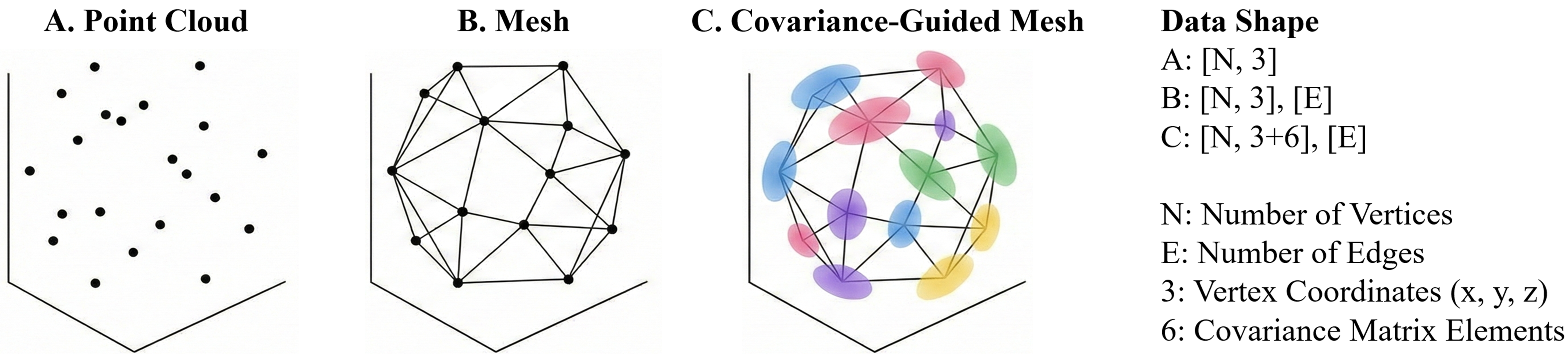
Figure 1: Schematic illustration of different mesh representations, highlighting gaps in correspondence and boundary fidelity.
Methodological Framework
HeartVolMesh advances cardiac mesh reconstruction via two synergistic modules:
Covariance-Guided Graph Deformation: Each template surface vertex is promoted to an anisotropic Gaussian kernel with learned mean and covariance, parameterized by a Cholesky factor for SPD stability. Vertex coupling is maintained via a mesh graph, and topology-consistent deformations are learned by a joint 3D CNN-GNN architecture. Covariance-aware negative log-likelihood loss provides direction-dependent geometric tolerance, crucial near ambiguous anatomical junctions.
Template-Driven Volumetric Warping: A volumetric tetrahedral template is warped to the reconstructed patient-specific surface through staged global alignment, non-rigid surface registration, and continuous deformation-field propagation. Connectivity and cross-case vertex/cell correspondence are preserved, with mesh resolution governed by template density.
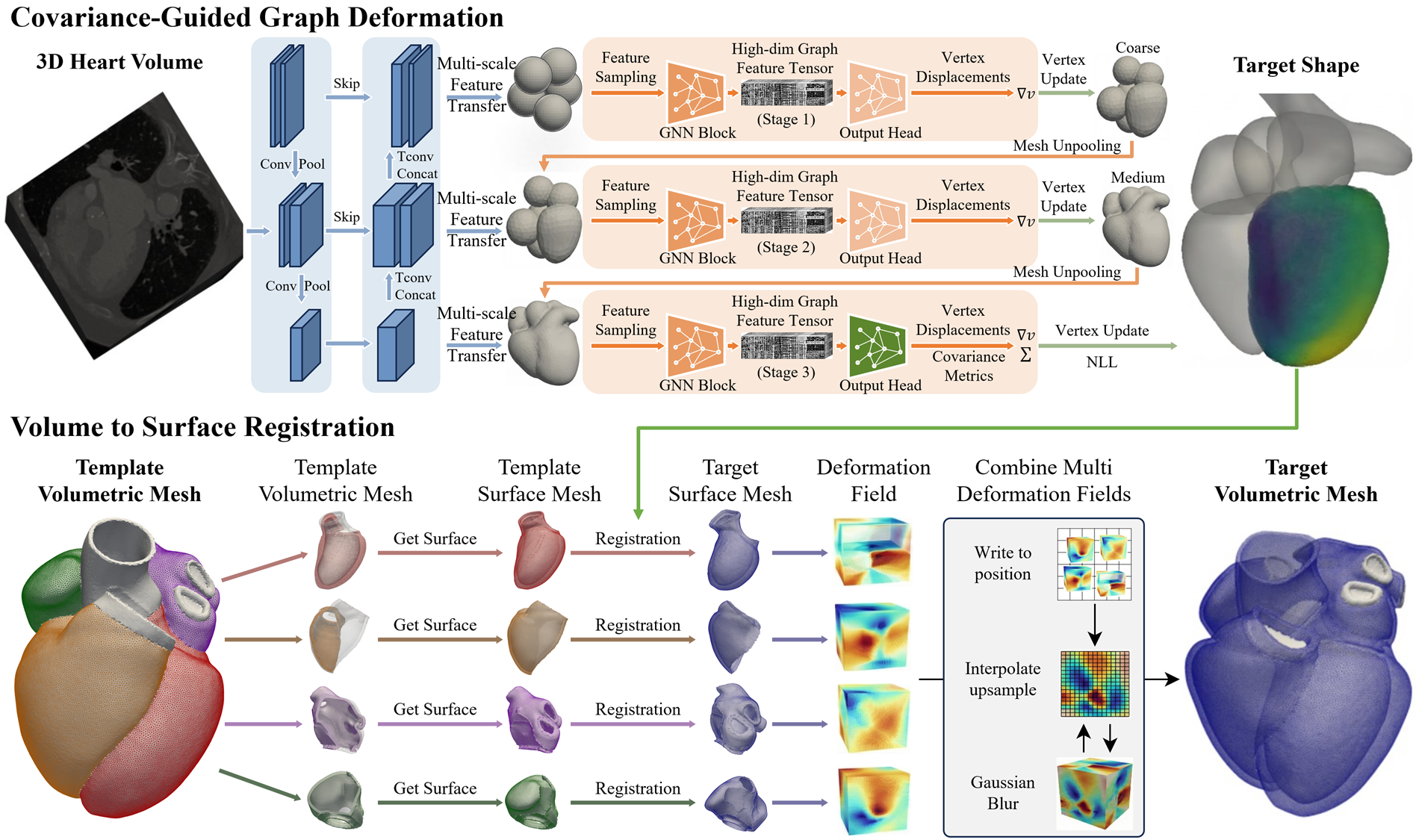
Figure 2: HeartVolMesh pipeline overview illustrating surface reconstruction via covariance-guided deformation, followed by template-driven volumetric mesh generation and warping.
Quantitative and Qualitative Evaluation
Experiments leverage a multi-centre dataset (900 patients, 4,000 temporal CTA volumes), utilizing a robust pseudo-GT benchmark derived from TotalSegmentator outputs with manual curation. Metrics encompass surface and volumetric mesh accuracy: Chamfer distance (CD), Hausdorff 95 (HD95), and normal consistency (NC), augmented with tetrahedral element quality statistics (minimum scaled Jacobian, minimum dihedral angle, inverted element count).
Volumetric Meshes: HeartVolMesh (Ours3p6) demonstrates superior boundary accuracy, achieving the lowest CD/HD95 values and highest NC across all volumetric targets, including thin-walled (LAMyo) and highly curved regions (LVMyo/RVMyo). The method yields 0.0% inverted elements, indicating strong simulation readiness.
Surface Meshes: Ours3p6 outperforms all baselines (including Ours3p1), evidencing the efficacy of anisotropic covariance modeling and supervision for resolving boundary ambiguity.
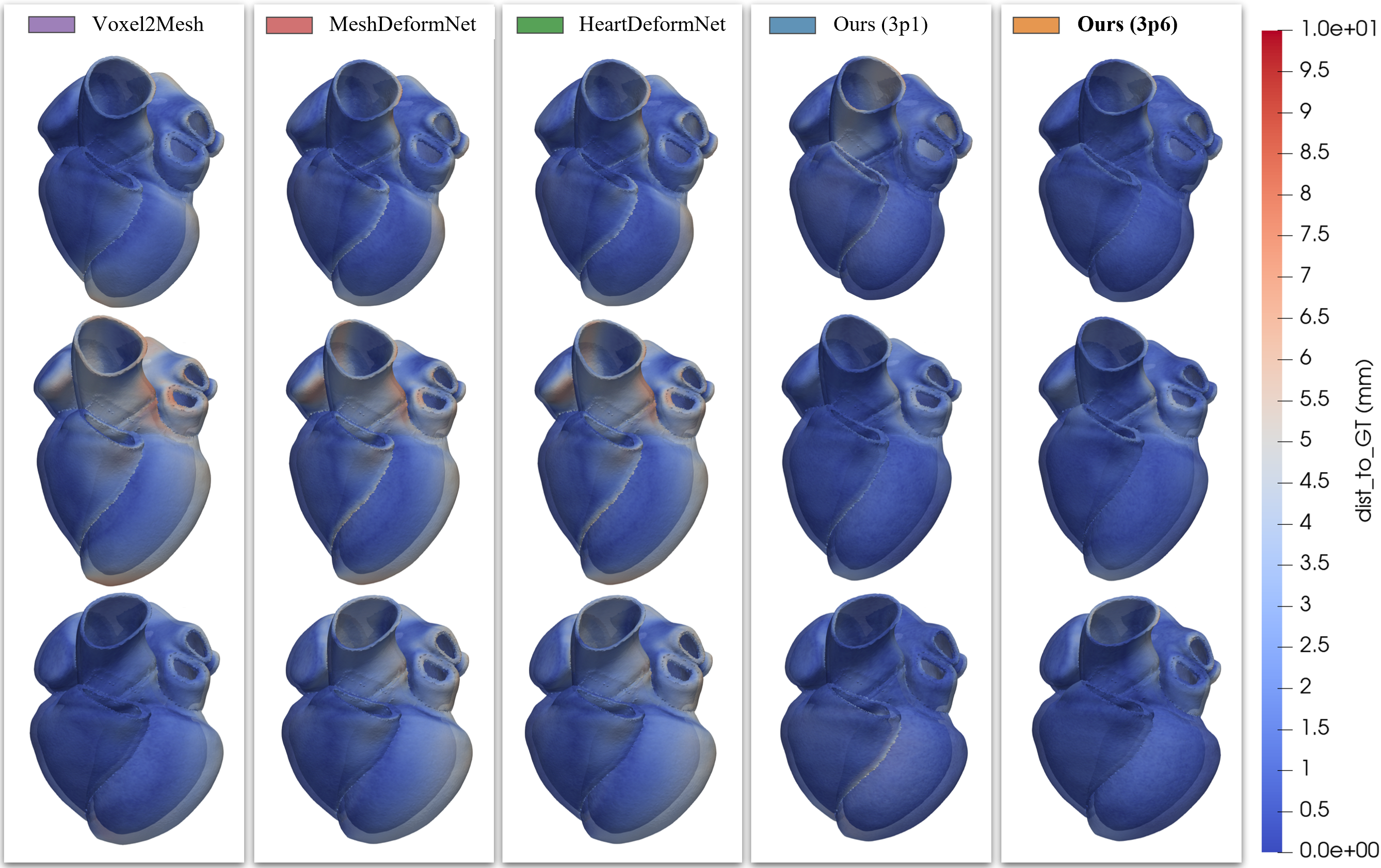
Figure 3: Qualitative volumetric reconstructions visualized for LA/LV/RA/RV/Myo; Ours3p6 exhibits sharper boundaries and reduced artifacts, especially at multi-structure junctions.
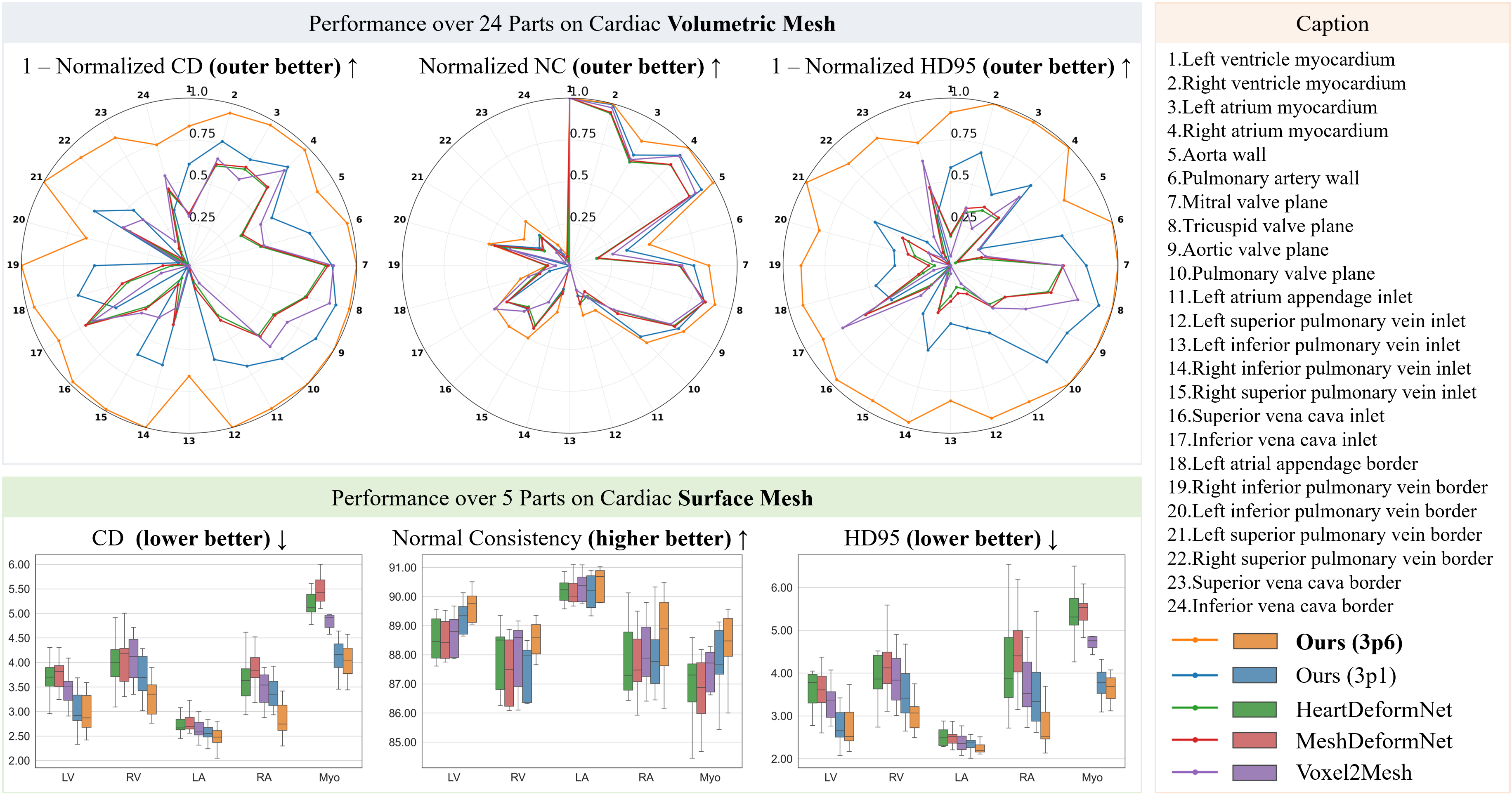
Figure 4: Quantitative summary: radar plots and boxplots of reconstruction metrics reveal that Ours3p6 achieves lower errors and reduced inter-case variance for both surface and volumetric structures.
Ablation Analysis
Three ablations dissect the methodological components:
- Ablation-A (Chamfer-only supervision): Degrades both surface and volumetric accuracy, highlighting the necessity of covariance-guided geometric matching.
- Ablation-B (coarse deformation-field): Mixed results; reduced deformation coherence oversmooths thin-wall regions, diminishing spatial accuracy.
- Ablation-C (no mesh regularization): Significantly worsens performance, demonstrating that regularizers (edge length, Laplacian, normal consistency) are essential for non-degenerate deformations and high-quality tetrahedral mesh propagation.
Practical and Theoretical Implications
HeartVolMesh directly addresses the gap in cross-case mesh correspondence and boundary fidelity, enabling scalable digital twin generation and population-level mesh analyses. The explicit incorporation of anisotropic uncertainty via vertex covariance facilitates robust handling of ambiguous junctions and thin-wall anatomy, critical in clinical imaging scenarios. The template-conditioned volumetric warping design accommodates flexible mesh density and element types, trading off fixed end-to-end meshes for adaptable simulation-focused specifications.
From a theoretical standpoint, the framework integrates probabilistic (Mahalanobis metric) and geometric (graph-based) mesh parameterization, aligning with modern uncertainty quantification practices. The decoupled surface/volume pipeline also respects anatomical priors and mesh topology constraints, paving the way for generalized mesh learning architectures.
Future Directions
Anticipated extensions include adaptation to cine CMR datasets, benchmarking with multiple template mesh specifications (density/element types), and direct simulation-based validations (electrophysiological, hemodynamic) to substantiate mesh fidelity for cardiac digital twin deployment. Furthermore, ongoing methodological advances in uncertainty-guided graph deformation may facilitate applications across other multi-structure organ systems and non-cardiac domains.
Conclusion
HeartVolMesh introduces a covariance-aware, template-driven paradigm for high-fidelity cardiac volumetric mesh reconstruction, achieving consistent improvements in boundary localization, volumetric correspondence, and element quality. Its principled design bridges the simulation-oriented requirements of in-silico trials with robust, population-scale digital twin construction, marking a methodological advance in medical image-to-mesh learning (2607.04243).