- The paper introduces a unified framework that reconstructs temporally resolved 4D whole-heart meshes directly from routine multi-view cine MRI.
- It employs multi-scale temporal modeling with a mesh VAE and physics-inspired differentiable contour rendering to achieve physiologically plausible motion and precise anatomical fidelity.
- The framework demonstrates significant improvements in reconstruction accuracy and stability, supporting downstream electrophysiological simulations and digital twin applications.
Personalized 4D Whole-Heart Mesh Reconstruction: Multi-Scale Temporal Modeling and Differentiable Contour Rendering
Introduction and Motivation
Personalized in-silico cardiac modeling, especially for cardiac digital twins (CDTs), requires anatomically and temporally accurate whole-heart reconstructions from routine clinical imaging. Despite the ubiquity of cine MRI for cardiac motion assessment, its sparse 2D multi-view nature (short-axis and long-axis slices) impedes robust 4D (3D+t) mesh-based reconstructions essential for biophysically faithful simulations. Traditional pipelines are limited by segmentation-to-shape routines, static geometry assumptions, or narrow anatomical coverage, frequently failing to capture physiologically coherent, patient-specific dynamics.
This work presents a unified, end-to-end framework that reconstructs temporally resolved 4D whole-heart meshes directly from routine multi-view cine MRI, combining image-to-mesh latent mapping, multi-scale temporal dynamics modeling, and a new physics-inspired differentiable rendering loss to enforce anatomy-aware supervision via multi-view slice contours.
Methodology
The proposed pipeline comprises several specialized modules:
- Domain-Specific Encoders: CMR U-Net for anatomical feature extraction from multi-view cine MRI and a mesh variational autoencoder (VAE) encoding both cardiac geometry and temporal mesh sequence priors.
- Image-to-Mesh Latent Mapping: Cross-domain mapping aligns cine MRI–derived anatomical embeddings with mesh sequence latent space, leveraging LoRA-modified mesh decoders for efficient, subject-specific mesh trajectory inference.
- Multi-Scale Temporal Modeling (MST): Dual-branch global (self-attention) and local (sliding window) temporal modeling ensures both cycle-level context and inter-frame coherence, crucial for smooth and physiologically plausible inter-chamber dynamics.
- Differentiable Contour Rendering: A Beer-Lambert law–inspired differentiable loss transforms mesh-to-plane distances into soft contour associations, enabling dense, plane-specific, anatomy-guided optimization from sparse 2D cine segmentations. This directly penalizes misalignment between projected reconstruction contours and manual segmentations across views.
- Geometric Regularization: Edge-length and normal-consistency terms stabilize mesh topology and local surface orientation.
Results
Anatomical and Kinematic Fidelity
The model outperforms baselines—especially HybridVNet—on whole-heart and sub-chamber MAE, with a reported overall whole-heart MAE of 1.68±0.31 mm and significant improvements in temporal smoothness (motion jitter 0.77±0.17 mm/frame3). Ventricular segments benefit from denser SAX sampling, while atrial localization remains more error-prone due to sparser long-axis coverage, quantitatively confirming known MRI protocol asymmetries.
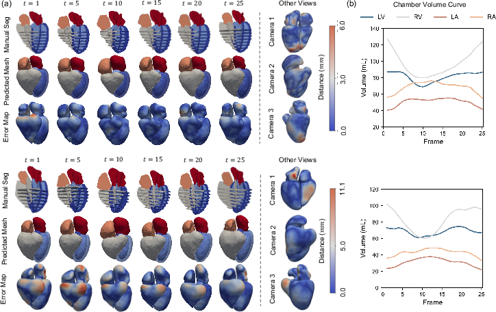
Figure 2: Illustration of whole-heart reconstruction quality from two representative cases at various cardiac phases, alongside the corresponding reconstructed chamber volume curves.

Figure 4: Chamber volume curves (mean ± std) for ground truth and predicted meshes; MST modeling improves spatiotemporal consistency.
Temporal Consistency and Function
The framework yields temporally coherent, physiologically plausible chamber volume curves closely matching reference trends. On post-MI populations, blunted LV volume excursions are accurately captured, with strong correlations in LVEDV, RVEDV, LVESV, RVESV, LVEF, and RVEF (r values >0.77), validating the utility of MST modeling for functional assessment.
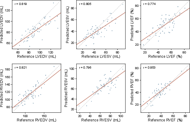
Figure 1: Correlation plots between predicted and reference ventricular functional indices. Both volumetric and functional metrics show high concordance.
Contour Alignment and Supervisory Efficacy
Direct boundary supervision through differentiable contour rendering improves alignment in both SAX and long-axis planes. The model achieves boundary F-score and mean contour distance metrics superior to variants lacking rendering loss. Visual assessment shows robust multi-view contour overlap, especially in complex anatomical regions.
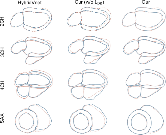
Figure 3: Multi-view qualitative comparison showing superior contour alignment when using the differentiable rendering loss.
Simulation-Readiness: Whole-Heart Electrophysiology
Meshes produced by the framework can be feasibly converted for in-silico electrophysiological (EP) simulations. Epicardial sequence, ventricular activation maps, and synthesized ECG signals derived from the reconstructed meshes conform with known physiological patterns, demonstrating the practical integration with downstream cardiac computational models.
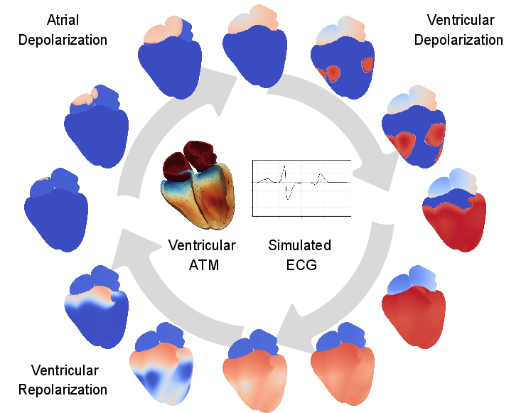
Figure 5: Whole-heart EP simulation output—action potential propagation, activation-time maps, and synthetic ECG based on reconstructed anatomy.
Ablation and External Validation
Ablation studies show all core components—multi-view fusion, U-Net pretraining, mesh VAE prior, MST block, and DR loss—are necessary for optimal reconstruction. Omission of any view, especially the 4CH plane, degrades performance markedly. MST modeling is critical for volumetric accuracy and motion plausibility.
External validation on the BAAI Cardiac Agent dataset with mixed vendors and missing views confirms robust generalization. LoRA adaptation improves cross-domain performance across Chamfer/HD, MAE, MSE, and jitter metrics, demonstrating resilience to imaging heterogeneity.
Implications and Future Directions
This work substantiates the feasibility of using routine multi-view cine MRI for direct, temporally resolved whole-heart mesh reconstruction, supporting both downstream EP simulation and integrated CDT workflows. The integration of differentiable rendering grounded in physical principles and MST latent dynamics sets a new standard for anatomical and functional consistency in sparse-supervision regimes.
Practical implications include rapid 4D mesh phenotype extraction for computational cardiology, functional quantification, and simulation-based risk stratification. Theoretically, the work bridges physics-informed supervision, spatiotemporal representation learning, and efficient latent adaptation for cross-modal medical image computing.
Future research will involve relaxation of explicit contour annotations, incorporation of biomechanical priors for tissue property estimation, extension to arrhythmic and non-sinusoidal cycles via sequence modeling, atlas-free topology adaptation, and validation on heterogeneous, multi-pathology populations. Joint optimization strategies—potentially through end-to-end graph networks or implicit neural representations—could further streamline segmentation, reconstruction, and simulation.
Conclusion
The approach delivers state-of-the-art accuracy and temporal coherence in 4D personalized whole-heart mesh reconstruction from routine clinical cine MRI, with demonstrated applicability to digital twin modeling and computational simulation. The framework addresses both the anatomical and kinematic requirements of CDT applications, indicating a viable path toward automated and scalable cardiac phenotyping in real-world clinical and research settings (2607.01952).