- The paper introduces a dual-branch implicit neural representation that fuses coordinate-based and image feature embeddings to enhance unsupervised cardiac cine MRI reconstruction.
- It leverages advanced positional encodings with complex-valued activations and iterative conjugate gradient refinement to improve both spatial and temporal image fidelity.
- Experimental results reveal that the method outperforms traditional techniques in PSNR and SSIM, demonstrating robustness under various sampling strategies and coil configurations.
Dual-Branch Implicit Neural Representations with Image Feature Embedding for Unsupervised Cardiac Cine MRI Reconstruction
Introduction and Background
Cardiac cine MRI is central to dynamic cardiovascular imaging, but accelerating data acquisition remains challenging due to SNR loss and artifact-prone reconstruction at high undersampling factors. Traditional methods, including parallel imaging and compressed sensing, are limited in achievable acceleration and often require sophisticated priors or expensive fully-sampled acquisitions. Deep learning-based methods have shown diagnostic-grade results in supervised settings but their reliance on fully-sampled references and large datasets restricts clinical translation.
Implicit Neural Representations (INRs) represent a promising direction—offering data-driven priors via compact continuous mappings from coordinates to signal values, and enabling scan-specific unsupervised reconstructions. Prior INR approaches, whether operating in k-space or image domain, typically rely exclusively on coordinate-based signal interpolation, underutilizing neighborhood context and limiting recovery in ill-conditioned settings. The paper "[Enhancing Implicit Neural Representations with Image Feature Embedding for Unsupervised Cardiac Cine MRI Reconstruction]" introduces I-FP-INR, a dual-branch image-domain INR model that integrates both coordinate and contextual image feature embeddings within an unsupervised coil-aware MRI reconstruction pipeline. This design addresses the aforementioned limitations by leveraging complex-valued feature extraction and iterative inference refinement to enhance spatial and temporal fidelity.
Methodology
Model Architecture
I-FP-INR comprises two synergistic branches:
- I-P-INR (Coordinate-based branch): Implements a network that accepts spatial-temporal coordinates (i,j,t) mapped via high-dimensional positional encodings (NeRF for spatial, Fourier features for temporal) into an MLP utilizing complex Gabor (WIRE) activations, optimizing for both high- and low-frequency detail.
- I-F-INR (Feature-based branch): Processes image feature embeddings extracted from current cine reconstructions using a complex-valued U-Net architecture. These features capture local and global context, and are embedded into an MLP (ReLU activations) yielding a complementary imaging signal.
Bidirectional interaction modules are present before and within both branches, facilitating information exchange at multiple integration points. The outputs of both branches are averaged and then data consistency is enforced through a conjugate gradient step.


Figure 1: The optimization pipeline of I-FP-INR, highlighting multi-coil and coil-combined strategies, sharing the dual-branch INR backbone with iterative inference refinement and data consistency enforcement.
Optimization and Loss
The reconstruction objective enforces consistency between reconstructed coil images and acquired k-space data. The loss comprises NIK (magnitude-focusing, regularized HDR loss) and relative L2 (phase-aware) terms, with location-adaptive weighting to mitigate dynamic-range disparities across k-space. The optimization alternates between network parameter updates and inference refinement stages—where reconstructed estimates are cyclically improved via data-consistent conjugate gradients.
Reconstruction Strategies
I-FP-INR is implemented under two strategies:
- Multi-coil: The network directly reconstructs all coil images, handling inter-coil encoding and sensitivity interactions jointly.
- Coil-combined: Reconstruction operates on coil-combined images, with expansion to coil space handled by physical constraints (coil sensitivity maps) during data consistency enforcement. This strategy tends to be more robust under sparse or irregular k-space sampling.
Experimental Evaluation
Datasets and Protocols
Experiments are conducted on the CMRxRecon2024 public dataset (330 subjects, multiple contrasts and sampling patterns, gold-standard for cine MRI), as well as in-house LUMC 0.6T body array data (short-axis bSSFP, variable- and fixed-density masks). Evaluations span common sampling schemes: Gaussian Cartesian (with/without ACS), VISTA, and Poisson disk, at acceleration factors 4 and 8.
Quantitative Results
I-FP-INR systematically outperforms strong baselines—including L+S, P-INR, KP-INR, Hash-INR, and single-branch I-P-INR—in both PSNR and perceptual metrics (SSIM, DISTS, HaarPSI), especially at higher R and under non-standard sampling. Notably:
- Under Gaussian Cartesian with ACS (R=4, R=8): I-FP-INR (multi-coil) achieves the highest fidelity, e.g., PSNR 45.11±2.20 (R=4).
- Under VISTA and PDS patterns (where central k-space is not fully sampled), coil-combined I-FP-INR demonstrates superior stability and artifact suppression, with strong effect size and statistical significance.
- Across both datasets, the dual-branch structure consistently delivers improvements over coordinate-only baselines, especially visible in temporal dynamic recovery and spatial coherence.
Qualitative Analysis
Visualizations reveal that I-FP-INR yields reconstructions with sharper features, fewer artifacts, and better preservation of cardiac structure across all tested sampling strategies and acceleration factors.
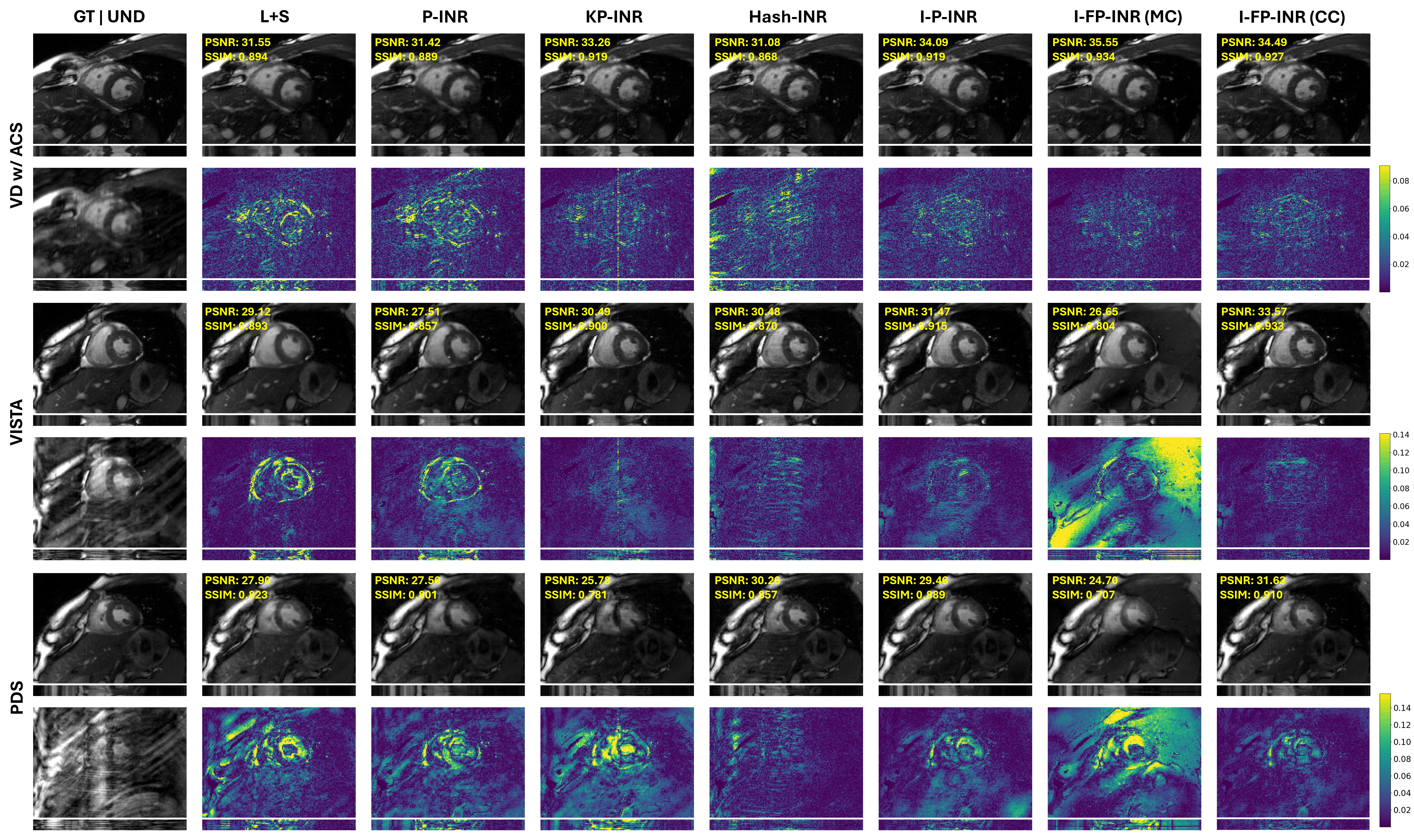
Figure 2: Qualitative comparison of I-FP-INR and baselines at R=4, all sampling patterns. Note restored sharp cardiac features and more accurate temporal dynamics in the dual-branch method.
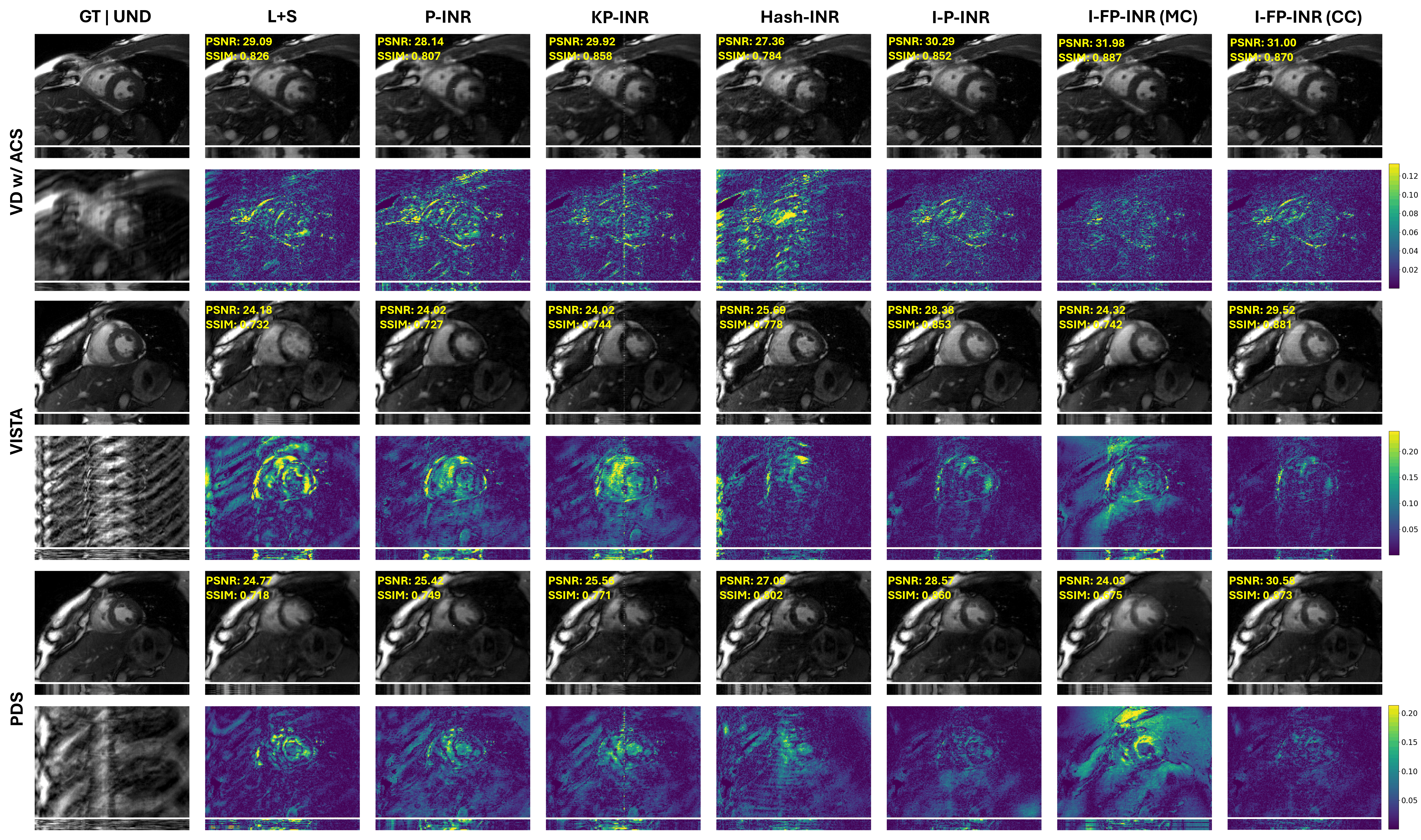
Figure 3: Qualitative comparison at R=8, demonstrating increasing importance of feature embeddings under aggressive undersampling.
Generalization to In-house Data
Performance on the LUMC in-house dataset reinforces robustness: both I-FP-INR strategies outperform classical L+S in fixed mask settings; the coil-combined variant maintains performance under frame-varying mask patterns at higher acceleration factors.
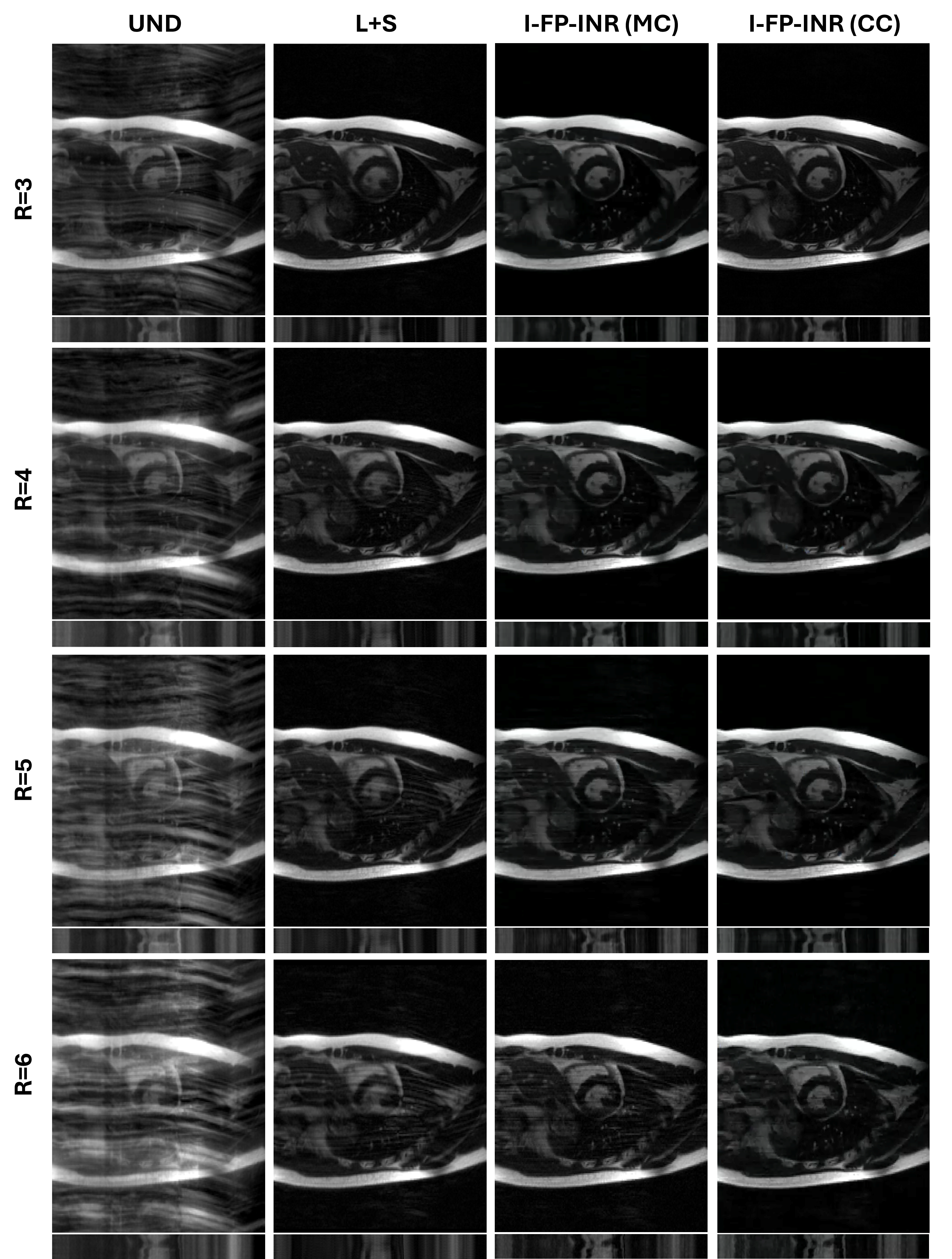
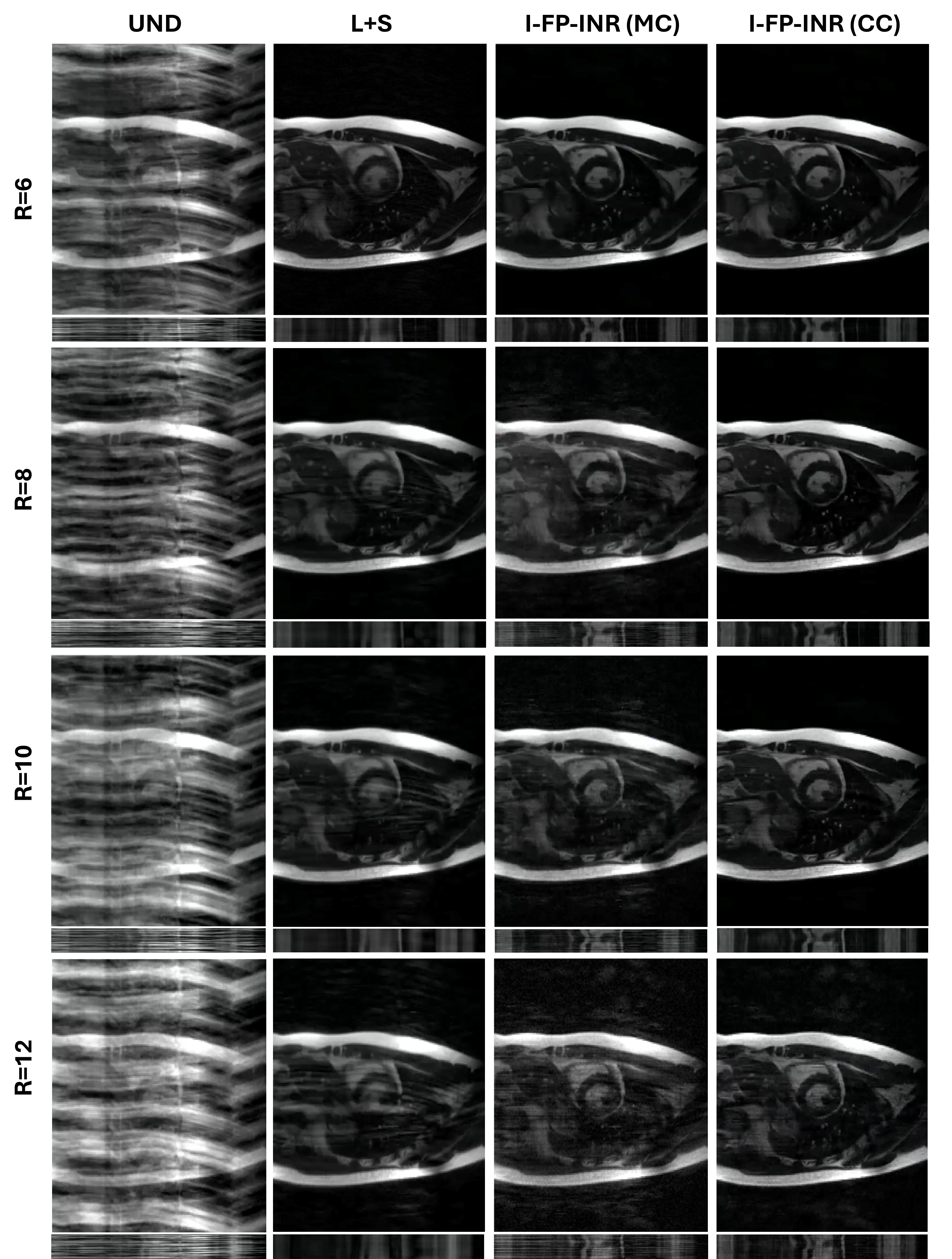
Figure 4: Visualizations from in-house data—temporal profiles indicate substantial suppression of artifacts and sharper structures, especially for I-FP-INR (CC) at extreme accelerations using dynamic masking.
Ablation Studies
- Branch composition: WIRE activation in the coordinate branch and ReLU MLP in the feature branch yield optimal results.
- Encoding: NeRF-based positional encoding for spatial and Fourier for temporal coordinates provide the highest fidelity.
- Loss composition: The combination of NIK and relative L2 losses is required for balanced magnitude-phase recovery.
- Inference stage: Removal consistently degrades performance, highlighting the importance of iterative refinement.
Implications and Future Perspectives
I-FP-INR establishes a new paradigm for unsupervised MRI reconstruction by integrating coordinate-based and feature-based implicit representation learning. The dual-branch design enhances information richness and stabilizes learning under severe undersampling and complex sampling patterns. Practical implications include:
- Improved diagnostic reliability for accelerated cardiac imaging with substantially reduced need for fully-sampled reference data.
- Strong robustness to vendor, field strength, sampling trajectory, and acceleration parameters.
- Potential extensibility to other dynamic imaging problems and domains requiring continuous signal reconstruction from sparse, non-uniform measurements.
The primary limitation is computational overhead, largely attributable to iterative feature extraction and inference refinement. Future work could investigate more hardware-efficient feature branches, adaptive iteration strategies, or transfer learning to amortize per-case computational burden. Furthermore, fine-grained modeling of physical acquisition processes and integration with physics-informed priors could yield additional fidelity gains.
Conclusion
I-FP-INR represents a notable advancement in unsupervised cine MRI reconstruction, leveraging dual-branch implicit neural representations that tightly couple coordinate mapping with contextual feature embeddings. The method achieves consistent improvements over state-of-the-art baselines in both coil-aware and coil-combined paradigms, especially under challenging sampling regimes and acceleration. These results point toward practical deployment of INR-type models in accelerated clinical cardiac MRI, and open avenues for cross-domain applications of dual-branch continuous learning frameworks.
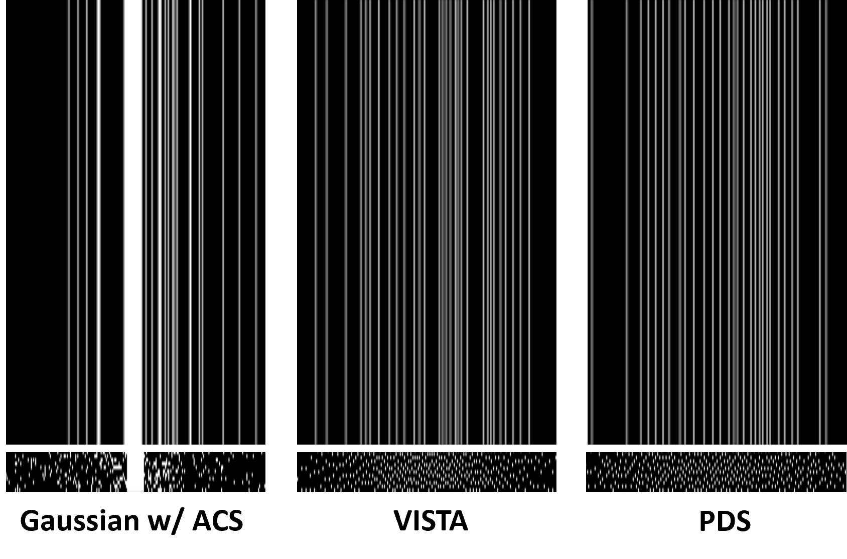
Figure 5: Examples of tested sampling masks at R=8 (top: k-space masks, bottom: corresponding temporal sampling profiles).