- The paper presents a novel SA-RDM-DC approach integrating residual generative drifting, hard data consistency, and self-auditing for improved MRI reconstruction accuracy.
- It demonstrates superior pathology preservation with enhanced SSIM, ROI-based metrics, and reliable error maps, ensuring clinical viability.
- The method achieves rapid, subsecond inference while maintaining robust performance under protocol shifts for effective selective review.
Self-Auditing Residual Drifting for Pathology-Preserving Accelerated Knee MRI: An Expert Analysis
Introduction and Motivation
Accelerated MRI reconstruction from undersampled k-space remains a key technical challenge, especially in anatomical regions like the knee where high-frequency pathology detail and small spatially localized abnormalities are clinically relevant. While deep learning-based models (e.g., U-Net variants, MoDL, VarNet) have demonstrably improved global fidelity metrics for MRI reconstruction, global metrics such as SSIM, PSNR, and NMSE inadequately capture clinically significant failure modes such as missed or distorted pathology regions, especially under high acceleration or protocol shift. The paper “Self-Auditing Residual Drifting for Pathology-Preserving Accelerated Knee MRI” (2607.02428) presents SA-RDM-DC, a novel architecture that synthesizes (1) a residual generative drifting paradigm with (2) explicit physics-based data consistency and (3) self-auditing reliability outputs to address these issues.
Methodological Overview
The core contribution of SA-RDM-DC is an integrated approach to MRI reconstruction that learns a residual-domain drift field, enforces hard measured-data consistency, and simultaneously performs self-auditing of reconstruction reliability in a feed-forward manner.
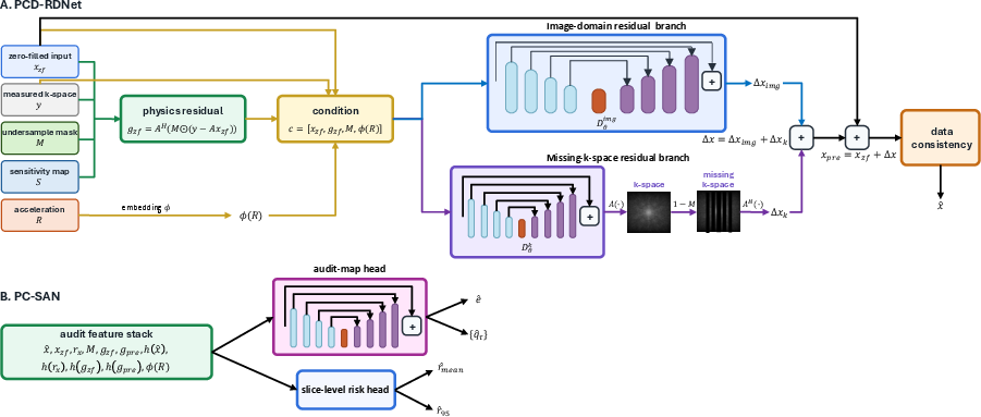
Figure 1: Overview of the SA-RDM-DC framework highlighting dual-residual drifting, data consistency, and concurrent prediction of dense error maps and risk scores.
PCD-RDNet: Physics-Conditioned Dual-Domain Residual Drifting Network
PCD-RDNet operates by learning a drift vector in the residual space from a zero-filled SENSE reconstruction toward the fully sampled residual correction. It comprises two branches: an image-domain residual branch and a missing-k-space residual branch. Both are conditioned on the zero-filled image, sampling mask, acceleration embedding, and physics-residual features computed as the measured k-space mismatch of the zero-filled input. A critical operation is the application of hard measured-k-space consistency, which strictly enforces fidelity to acquired data and controls hallucination.
Supervision integrates voxel-wise loss, SSIM, and a suite of frequency- and data-consistency-focused constraints. In the residual generative drifting loss, the predicted residual is attracted toward the fully sampled residual, with a conditional drift field guiding this generative transport. This approach mitigates the pathologies of both pure regression and unregularized generative sampling by aligning learned drift with the ground truth residual in measurement-consistent ways.
PC-SAN: Physics-Calibrated Self-Auditing Network
PC-SAN leverages the same physics-conditioned residual features to predict both dense error maps and slice-aggregated risk scores. The audit feature stack includes magnitude and high-pass images, residuals, k-space mismatch projections, and acceleration context. Dense error maps are optimized using a weighted loss that up-weights high-true-error regions, while slice-level risk scores use Huber losses for mean and tail (e.g., 95th percentile) error prediction. Additional quantile-regression heads provide calibrated pixelwise upper bounds on anticipated reconstruction error.
During inference, both the reconstruction and audit outputs are jointly produced, enabling rapid case-level triage and selective review.
Evaluation Protocol
Validation was carried out on fastMRI knee MRI and SKM-TEA datasets, employing metrics that holistically reflect both global image fidelity and clinical utility:
- Global metrics: SSIM, PSNR, NMSE, MAE, and high-frequency error norm (HFEN).
- Pathology-aware metrics: ROI-based SSIM, HFEN, edge preservation, and task-preservation via a frozen multi-label classifier for downstream pathology prediction stability.
- Self-auditing evaluation: Spearman correlation, AUROC, AUPRC of predicted risk scores against true reconstruction error, and risk-coverage (selective review) analysis.
- Protocol-shift (domain-shift) testing: Generalization and adaptation between fastMRI and SKM-TEA sequences.
Quantitative and Qualitative Results
Global and Pathology-Region Fidelity
SA-RDM-DC achieved the highest SSIM values across all tested acceleration factors (R=4,8,12), maintaining strong global fidelity even at high acceleration (see Table 1 in the manuscript). Notably, it preserves pathology-region structure more robustly than all competitive baselines, as measured by ROI SSIM, HFEN, and edge-error within radiologist-annotated fastMRI+ regions.
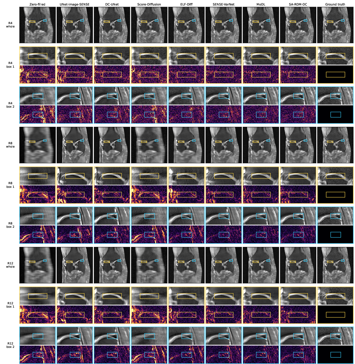
Figure 2: Visual comparison of reconstructions across acceleration factors, highlighting anatomical detail preservation in cartilage and meniscus regions by SA-RDM-DC compared to other baselines.
Runtime and parameter count benchmarks indicated that, unlike iterative diffusion and bridge models, the drift-based approach enables subsecond inference suitable for real-world deployment, without sacrificing critical detail.
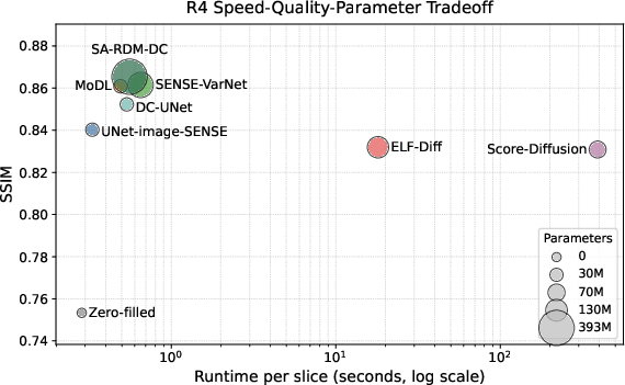
Figure 3: Trade-off plot between runtime, SSIM, and model size demonstrating SA-RDM-DC’s efficiency compared to sampling-based and unrolled methods.
SA-RDM-DC’s audit head exhibited near-ideal monotonicity and calibration with true slice-wise reconstruction error across all accelerations, achieving Spearman correlations >0.95 and AUROC >0.98 for detection of high-error slices.
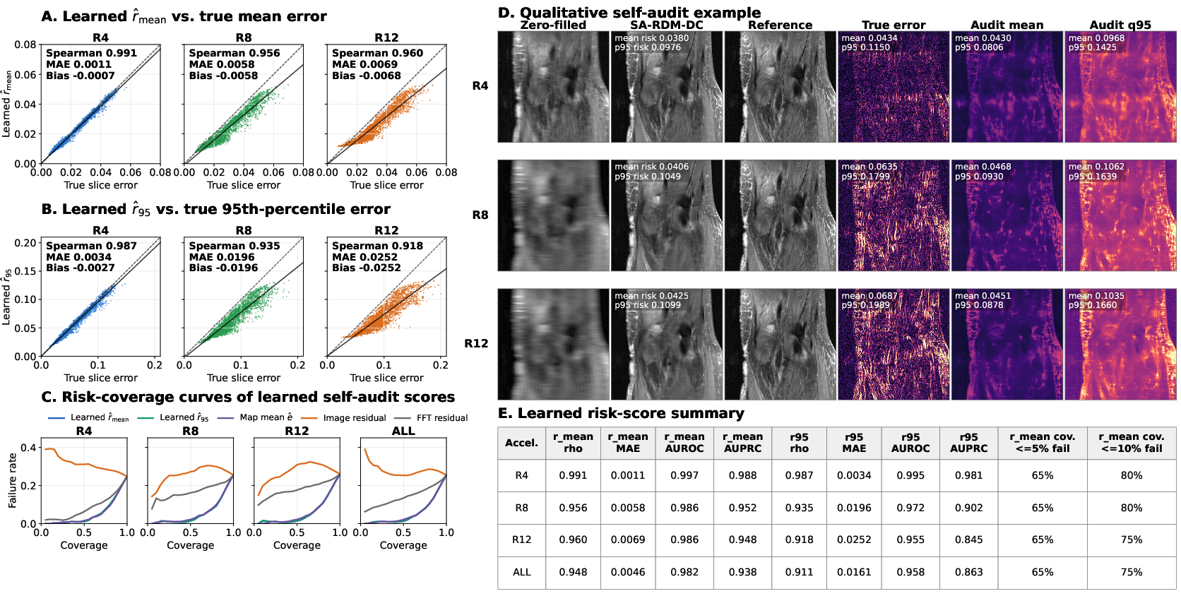
Figure 4: Self-auditing outputs including correlation plots, risk-coverage analysis, and qualitative error-map visualization. SA-RDM-DC audit head localizes error in anatomical context, outperforming naive residual baselines.
The audit head retained >75% coverage at ≤10% failure rate, underlining its practical capacity for selective review. Error maps and risk scores were visibly localized to unreliable regions rather than correlating merely with anatomical variations in signal.
Protocol Shift: Robustness and Adaptability
Zero-shot generalization to SKM-TEA data resulted in competitive SSIM and PSNR, outperforming most direct baselines (except MoDL in certain fine-tuned settings). Fine-tuning on SKM-TEA further improved metrics but highlighted necessary recalibration of audit models when applied to significantly different acquisition protocols.
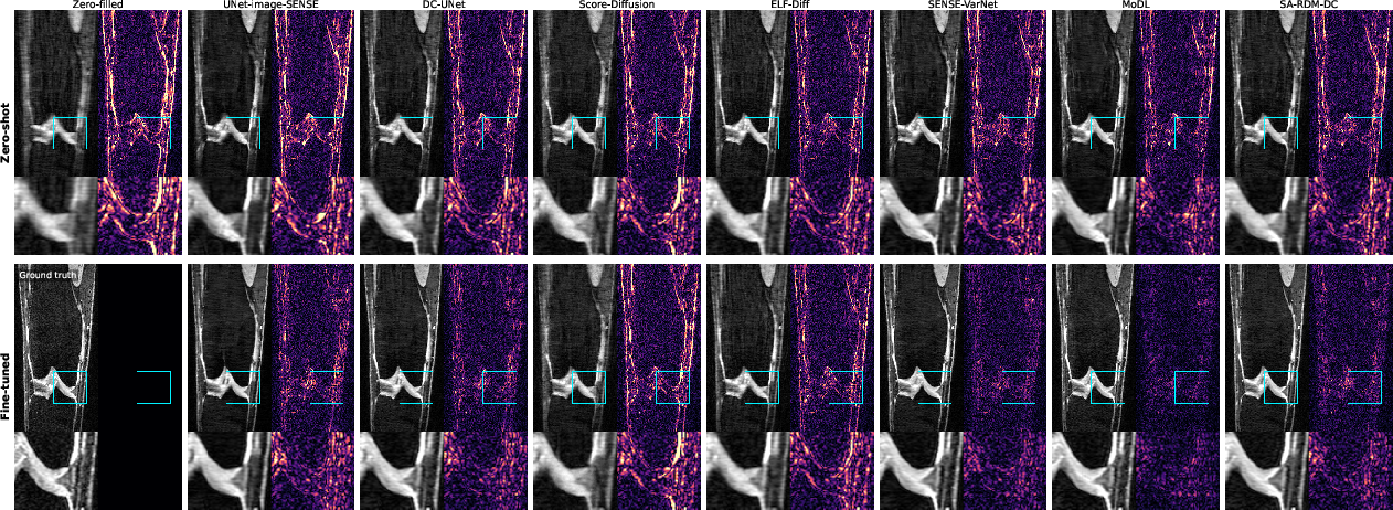
Figure 5: Protocol-shift qualitative reconstructions, illustrating domain-mismatch artifacts in zero-shot transfer and their mitigation after fine-tuning.
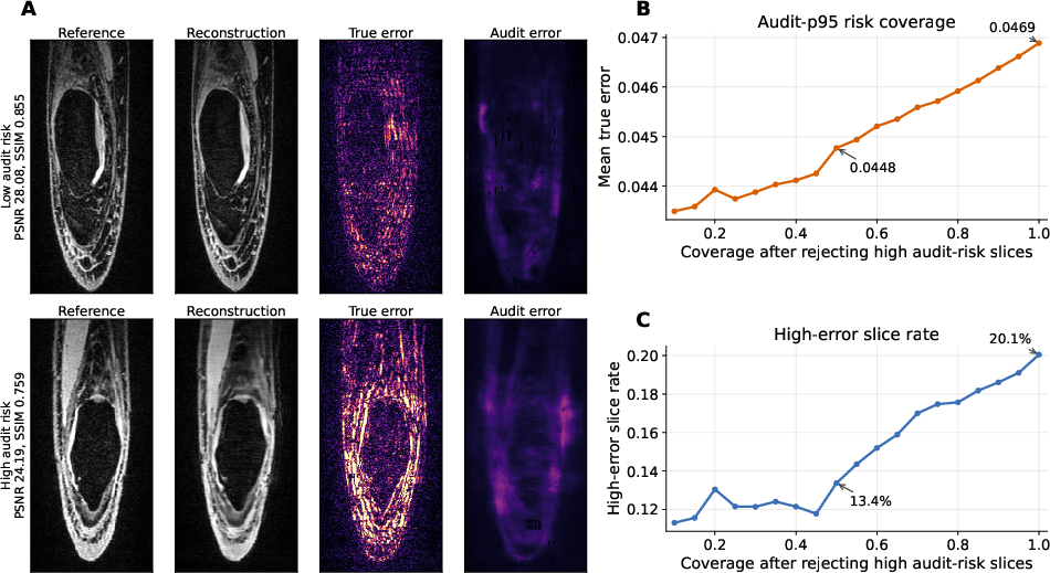
Figure 6: Post-fine-tuning self-audit behavior on SKM-TEA, showing partial transferability and the limits of calibration under protocol shift.
Ablation and Component Importance
Ablation studies validated the architectural integration:
- The removal of hard data consistency led to the most deleterious impact on all metrics, confirming its essential safeguarding role.
- Physics-based residual features and missing-k-space branches further contributed to improved detail recovery.
- The drift paradigm itself promotes structural fidelity in regions prone to high-frequency loss or nonuniform error.
Implications and Future Directions
SA-RDM-DC advances MRI reconstruction by demonstrating that residual generative drifting, when combined with explicit measured-data enforcement and learned reliability signals, enables high-fidelity, clinically robust reconstruction with actionable uncertainty outputs. The explicit audit head supports practical workflows where automated reconstructions are triaged for manual review, a necessity in many clinical and cross-protocol settings.
For future work, key directions include:
- Generalization analysis across broader anatomical sites, vendors, and acquisition protocols.
- Extension of pathology- and biomarker-based evaluation beyond bounding-box endpoints to segmentation-derived quantitative biomarkers.
- Calibration transfer and adaptation mechanisms for the audit head, including domain adaptation techniques and unsupervised reliability transfer across unseen domains.
- Integration of prospective radiologist reader studies for task-based and clinical-readout evaluations.
Conclusion
SA-RDM-DC establishes a compelling design point in deep MRI reconstruction: high-acceleration, pathology-preserving fidelity combined with feed-forward self-audit, realized through residual generative drifting and multifaceted physics guidance. Its evidence-based improvements in structural fidelity, meniscus prediction stability, and runtime efficiency mark it as an architecturally distinct and practically relevant advance for reliable, accelerated MRI reconstruction (2607.02428).