- The paper introduces a multimodal graph neural network that leverages hierarchical clinical priors and relation-aware attention for pCR prediction.
- It employs domain-adversarial decoupling to mitigate multicenter MRI protocol biases, achieving superior AUC and balanced accuracy compared to baseline models.
- The integration of an LLM-driven retrieval module enhances interpretability by providing evidence-based, post-hoc rationales for clinical decision-making.
ClinRAG-GRAPH: Clinical-prior Retrieval-Augmented Graph Model with Domain Adversarial Learning for Breast pCR Prediction
Introduction
Achieving robust, generalizable pre-treatment prediction of pathological complete response (pCR) under neoadjuvant chemotherapy (NAC) in breast cancer remains nontrivial due to high cross-modal heterogeneity and substantial domain shift induced by multicenter MRI protocol variations. The paper introduces ClinRAG-GRAPH, a multimodal graph neural network integrating DCE-MRI, structured clinical variables, and biopsy-derived pathological biomarkers using a hierarchical clinical-prior graph, prior-guided relational attention, domain-adversarial decoupling, and LLM-based retrieval-augmented inference. The study addresses documented limitations of classical fusion-based and neural approaches, including weak cross-modal dependency modeling, insufficient domain shift mitigation, and limited clinical interpretability. Methodological contributions are substantiated by comprehensive multicenter validation, ablation analysis, and interpretability assessments.
Methodology
Hierarchical Clinical-prior Graph Construction
ClinRAG-GRAPH encodes clinical knowledge via a directed intra-patient graph, where nodes correspond to imaging and tabular variables, and directed edge priors (puv) are explicitly stratified: guideline-level associations ("strong"), expert-agreed connections ("soft"), and weak/unproven dependencies ("learnable"). Edge priors modulate the first-layer attention weights, calibrating early message passing. This mechanism supplants ad hoc or fully data-driven connectivity, reducing the risk of semantic dilution and instability associated with auxiliary loss-based regularization. The approach is grounded in expert guidelines and radiologist consensus.
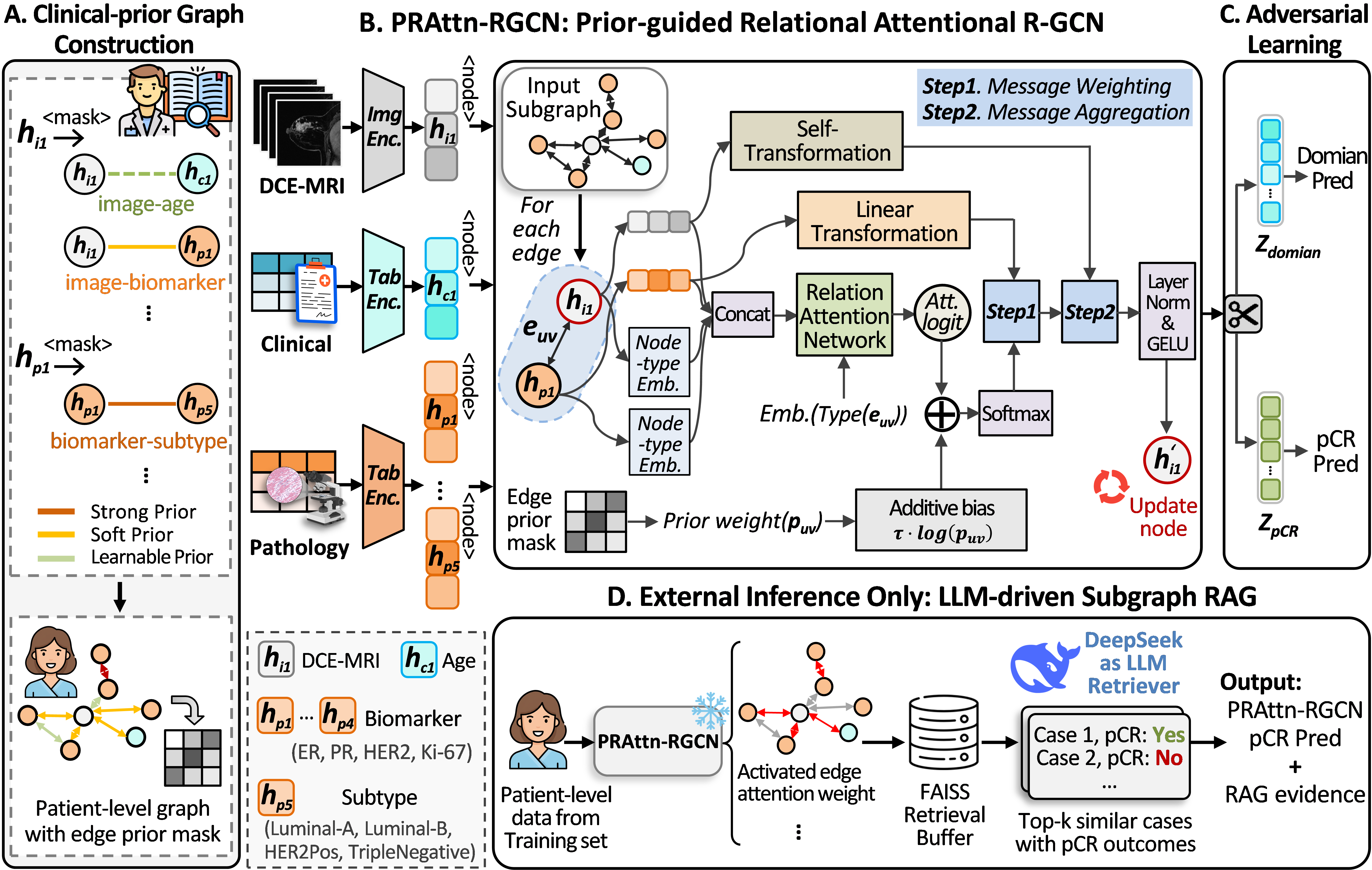
Figure 1: Overview of ClinRAG-GRAPH comprising graph construction, prior-guided R-GCN, adversarial decoupling, and retrieval-augmented inference.
Prior-guided Relational Attentional GCN (PRAttn-RGCN)
The message passing protocol leverages relation-type specific attention, with relation priors injected as additive log-biases at the input layer, thus integrating clinical hierarchy directly into the graph topology and information flow. PRAttn-RGCN operates in a hierarchical fashion: initial feature fusion is guided by clinical priors, but subsequent layers remain data-adaptive, balancing knowledge-driven structure and empirical optimization. The model readout incorporates both imaging and tabular (clinical, pathologic) representations aggregated via mean pooling.
Domain-Adversarial Decoupling
To mitigate multicenter MRI protocol bias, ClinRAG-GRAPH incorporates gradient reversal-based adversarial training on the MRI embedding. An auxiliary domain classifier with a GRL enforces center-invariant representations, promoting domain generalization without diluting predictive fidelity for pCR. Task and adversarial objectives are balanced via a trade-off parameter optimized empirically.
LLM-driven Subgraph RAG for Evidence-grounded Inference
A critical addition is the retrieval-augmented generation (RAG) submodule, driven by the DeepSeek LLM. Query patients are encoded, and subgraph-aware, schema-constrained search retrieves analogous historical samples using FAISS. Nearest neighbors are identified by combined representation and edge-wise attention signature similarity. Fused outputs from both the R-GCN and RAG provide final predictions. Structured rationales are auto-generated as post-hoc explanations, leveraging only retrieval metadata and subgraph matches—ensuring interpretability and evidence traceability in inference.
Experimental Evaluation
Multicenter Cohort and Implementation
ClinRAG-GRAPH is systematically evaluated on two public (DUKE, ISPY1) and three in-house datasets (“Zcenter”, “Ycenter”, “Qcenter”) with internal/external train-test splits established at the patient level to avoid information leakage. DCE-MRI inputs are tumor-centric, temporally concatenated volumes; clinical/pathological variables are standardized. Models are implemented in PyTorch and trained with AdamW, using careful batch sizing, normalization, and hyperparameter tuning for computational stability.
Comparative Analysis
Quantitative results demonstrate strong internal test AUC (0.815, 95% CI: 0.738–0.885) and robust external generalization, e.g., 0.774 AUC (Ycenter), exceeding R-GCN, LMF, iMRhpc, and M2Fusion. Notably, ClinRAG-GRAPH yields higher balanced accuracy and specificity compared to direct fusion and non-graph multimodal baselines. DeLong testing confirms statistical significance (p<0.05) of the model's superiority.
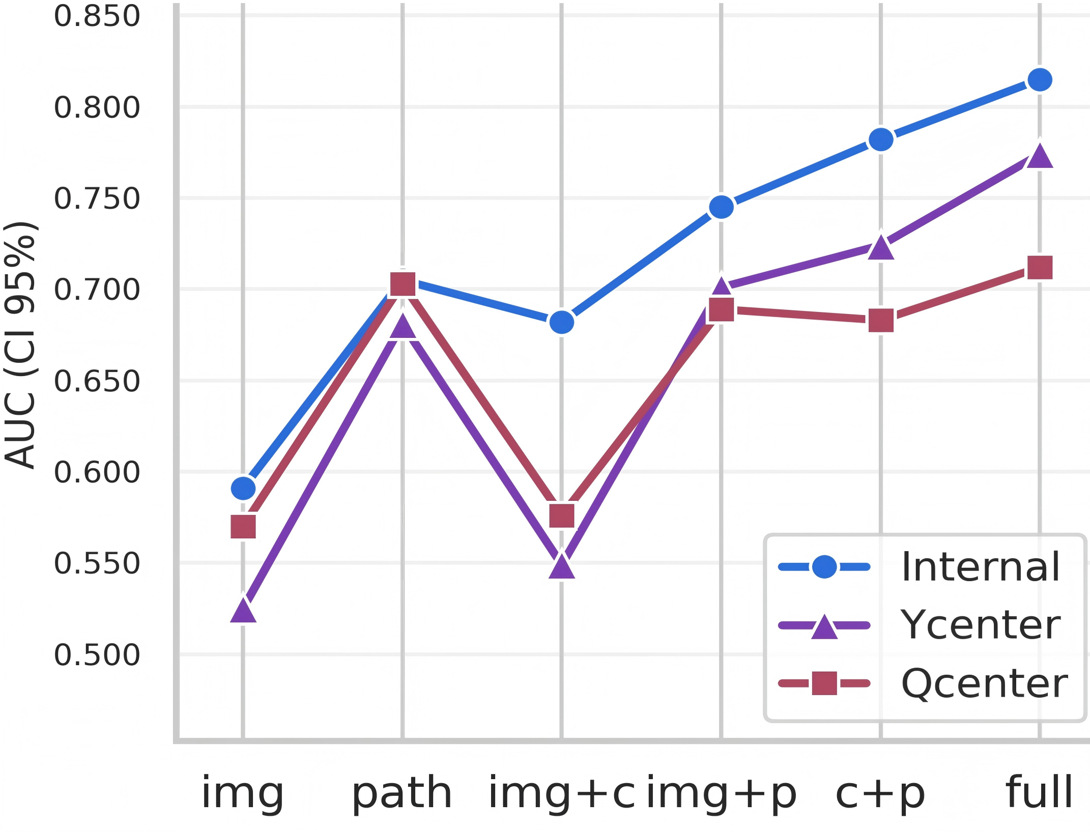
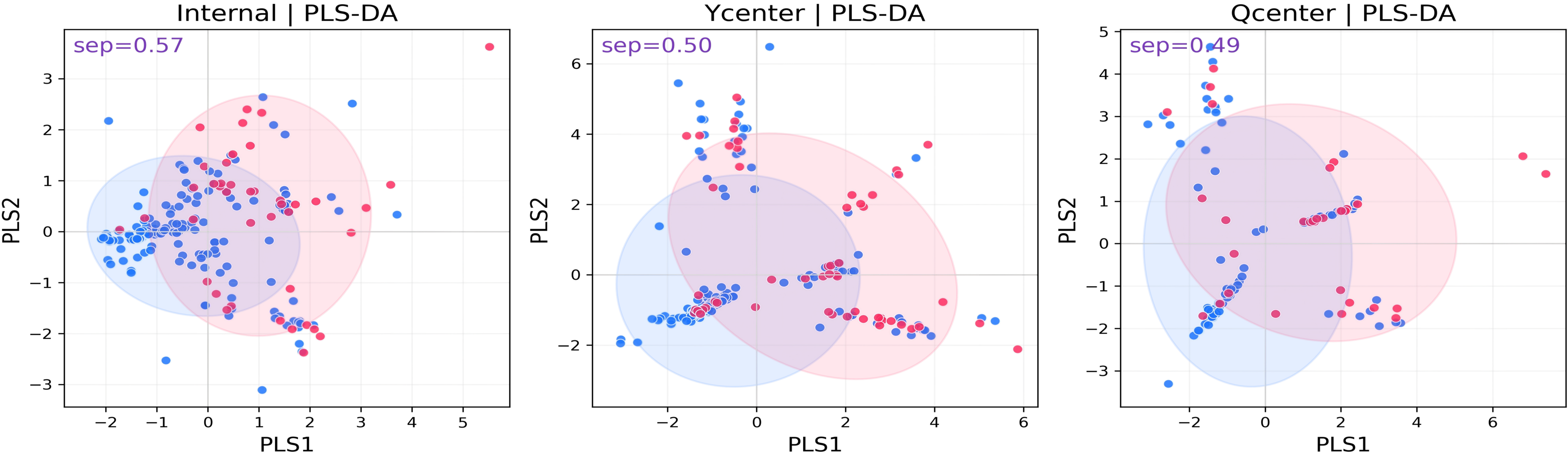
Figure 2: AUC for modality pairs, illustrating importance of pathology and multimodal fusion in model performance.
The model exhibits greater sensitivity for challenging, previously unseen Qcenter cases, underlining domain shift robustness. PLS-DA projections further reveal consistent class separation across internal and external test cohorts.
Ablation and Modality Contribution
A modular ablation study isolates the gains conferred by clinical-prior graphs, relation-aware attention, adversarial decoupling, and LLM-driven RAG. Each component incrementally improves AUC, with the complete pipeline yielding the highest scores. Modality-specific ablation verifies the dominant predictive power of pathology features, with DCE-MRI and clinical data providing complementary value.
Interpretability and Case Study
The internal mechanics of ClinRAG-GRAPH are interrogated using SHAP analysis, validating that attention-weighted graph edges map to biologically and clinically plausible interactions, especially across key molecular subtypes (Luminal A/B, HER2+, TripleNeg). Edges linking ER, PR, HER2, Ki67, age, and MRI retain high attribution values, indicating the network's reliance on structured dependencies over naive feature concatenation.
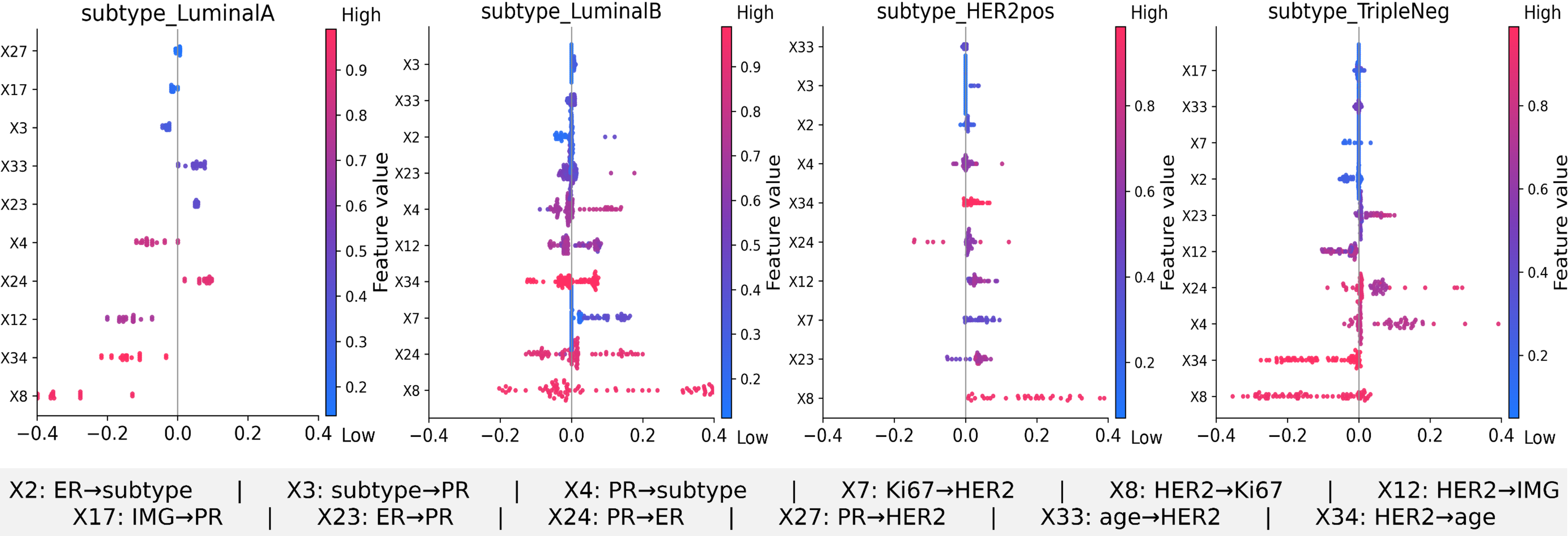
Figure 3: SHAP analysis: edge-level importance per molecular subtype, showcasing interpretable, subtype-specific relational reasoning.
A LLM-driven RAG case study demonstrates practical retrieval and explanation workflow for a patient (pCR=0, external set): DeepSeek identifies top-3 matched cases with highly fused similarity, rationalizes the decision via driver-edge patterns, and delivers a JSON-verifiable rationale, exemplifying the system's interpretability and auditability.
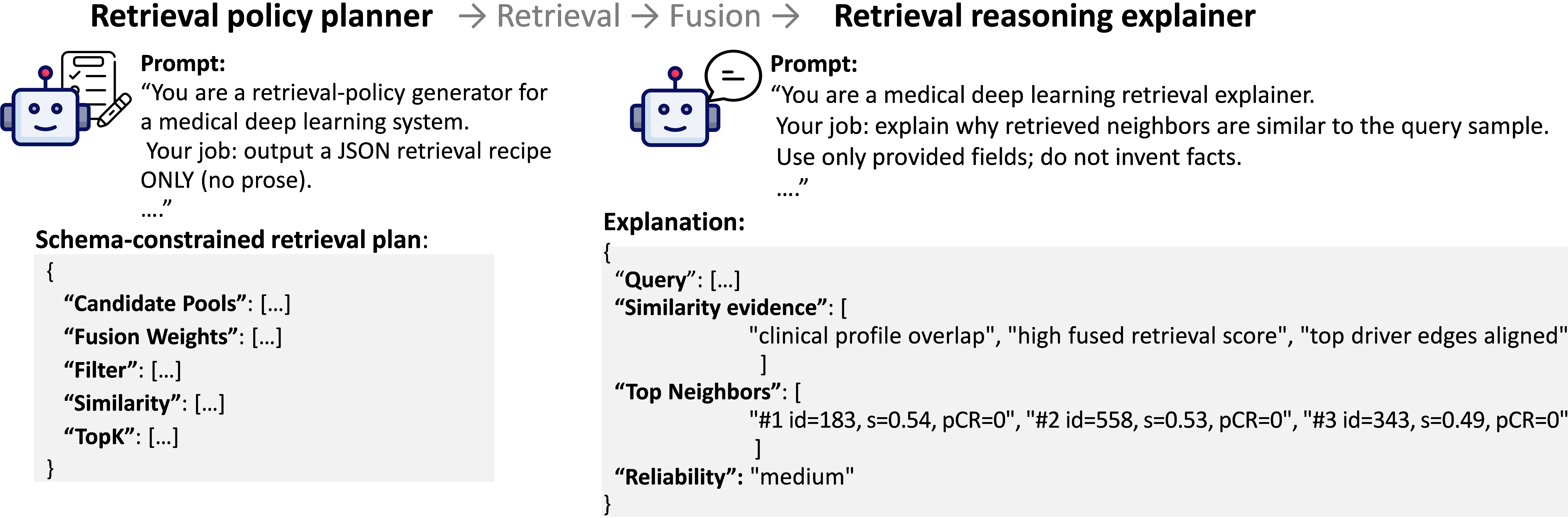
Figure 4: LLM-driven RAG deployment on an external cohort patient, illustrating evidence aggregation and result explanation.
Practical and Theoretical Implications
ClinRAG-GRAPH exemplifies an interpretable, multimodal AI pipeline capable of robust generalization under real-world heterogeneity and domain shift—key for prospective clinical translation of pCR prediction in multicenter settings. The explicit injection of clinical priors, modular adversarial decoupling, and LLM-augmented evidence reasoning delineate a paradigm for combining structured knowledge with data-driven learning. These principles could be extended to other oncologic or multimodal diagnostic domains requiring domain generalization and explainability.
The integration of large-scale LLMs as schema-constrained retrieval planners and post-hoc explainers introduces new opportunities for hybrid symbolic-neural inference in medical AI, subject to future advances in LLM transparency and clinical alignment.
Conclusion
ClinRAG-GRAPH advances the field of breast cancer pCR prediction by fusing hierarchical clinical knowledge graphs, relation-aware message passing, domain-adversarial learning, and RAG via LLMs—aligned for robust and interpretable multimodal inference. The pipeline outperforms conventional and state-of-the-art baselines in multicenter studies, and its interpretability mechanisms ensure clinical transparency. Ongoing extensions towards longitudinal and multi-timepoint modeling are poised to further enhance early therapy adaptation in oncology.