- The paper introduces a two-stage, disentangled framework that separates canonical volumetric inference from patient-specific deformation modeling.
- It achieves superior segmentation performance with metrics such as 83.83% Dice, enhanced SSIM, and PSNR, outperforming traditional methods.
- The method leverages dual-encoder fusion with adaptive cross-attention to integrate deep radiographic and semantic features for robust 3D reconstruction.
HyDAR-Pano3D: Disentangled Anatomical Recovery for Panoramic-to-3D Dental Reconstruction
Introduction and Motivation
The clinical dependence on panoramic radiograph (PR) imaging for screening and planning in dental practice contrasts sharply with its fundamental limitation: PR images produce a compressed 2D projection riddled with non-linear geometric distortion and depth ambiguity, failing to capture the true 3D complexity of oral structures. Direct 2D-to-3D reconstructions from PR data are thus extremely ill-posed, requiring the regressed model to recover both anatomical density and patient-specific spatial deformation, a conflation that induces over-smoothed, anatomically imprecise volumes.
To address these structural and inferential ambiguities, the proposed HyDAR-Pano3D framework departs from traditional monolithic 2D-to-3D regression, introducing a two-stage, disentangled paradigm. This architecture decouples the volumetric density estimation—performed in an anatomically normalized, "canonical" space—from patient-specific geometric restoration via deformation modeling. The result is a substantial reduction in learning ambiguity, with marked improvements in both volumetric fidelity and downstream clinical utility.
Methodological Framework
HyDAR-Pano3D operates via two sequential stages: canonical 3D anatomical inference and anatomical restoration.
Stage 1: Canonical Volumetric Inference.
A dual-encoder architecture is developed wherein deep radiographic features (extracted by the 3DPX encoder) are fused via adaptive cross-attention (ACF) with semantic priors from a domain-adapted vision transformer (SemiT-SAM). The ACF module aligns and integrates multi-scale feature pyramids from both radiographic and semantic backbones, modulating their influence via gated residual connections. This process leverages high-level anatomical priors inherent to contemporary foundation models to address the severe depth ambiguity and structural overlap characteristic of single PR projections.
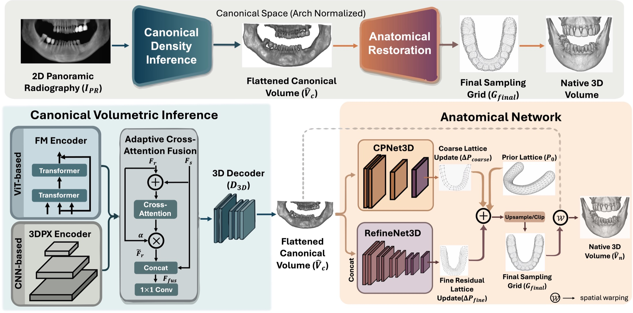
Figure 1: HyDAR-Pano3D factorizes 3D dental reconstruction into canonical volumetric inference (Stage 1) and anatomical restoration via deformation (Stage 2).
Stage 2: Anatomical Restoration Network (AR-Net).
After synthesizing a canonical arch-normalized 3D volume, a hierarchical, lattice-based deformation field is predicted. This field, initialized with an analytic dental arch prior, is estimated in a coarse-to-fine manner: a global transformation lattice captures large-scale arch morphology, whereas a residual refinement stage models localized patient-specific anatomical deviations. Differentiable backward resampling (warping) is then used to transform the canonical volume into the native patient space, restoring individualized morphology without confounding volume synthesis with spatial deformation.
Experimental Validation
The framework is evaluated on three diverse clinical CBCT datasets (NC, ST, TF), with test protocols involving synthetic PR volumes generated by curved planar reformation to enforce anatomical alignment for rigorous comparison.
Numerical results demonstrate substantial gains:
- On the TF dataset, HyDAR-Pano3D achieves 83.83% Dice, 85.70% SSIM, and 22.98 dB PSNR, a significant margin above state-of-the-art baselines.
- Improvements are consistent across challenging domains, with the method outperforming 3DPX and Oral-3D even amidst prevalent metal artifacts (TF and ST datasets).
- Downstream segmentation performance, as measured by Dice and HD95 for both whole teeth and the inferior alveolar canal (IAC), also improves, e.g., 82.4% Dice for teeth and 72.2% Dice for IAC on TF (an ~10% improvement over the strongest competing baseline).
Salient qualitative improvement can be observed in recovery of subtle dental structures, continuity of the mandibular canal, and preservation of tooth morphology, as demonstrated by segmentation overlays.
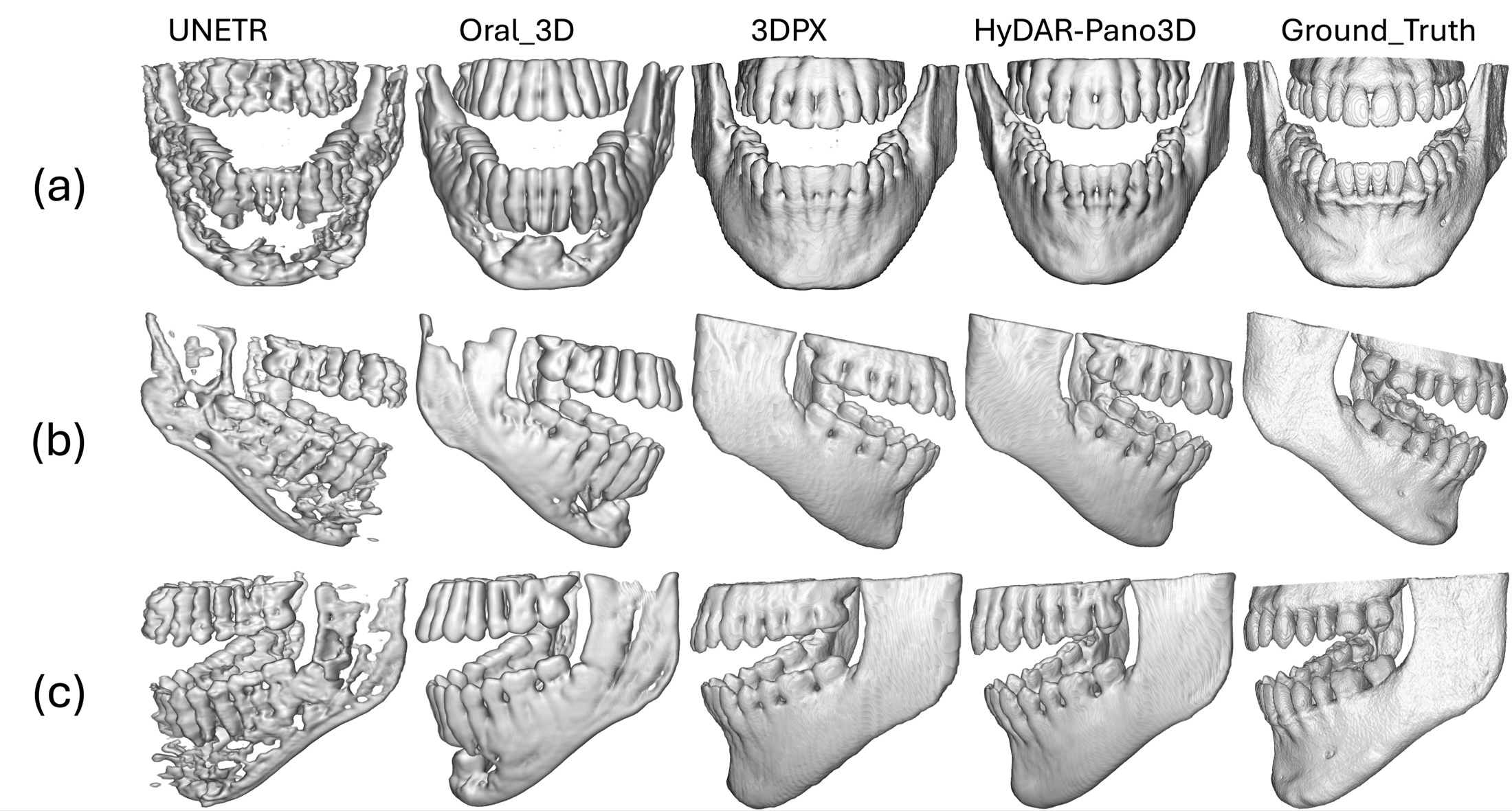
Figure 2: Qualitative comparison of panoramic 2D-to-3D dental reconstruction on TF cases; HyDAR-Pano3D maintains sharper anatomical fidelity versus competing models.
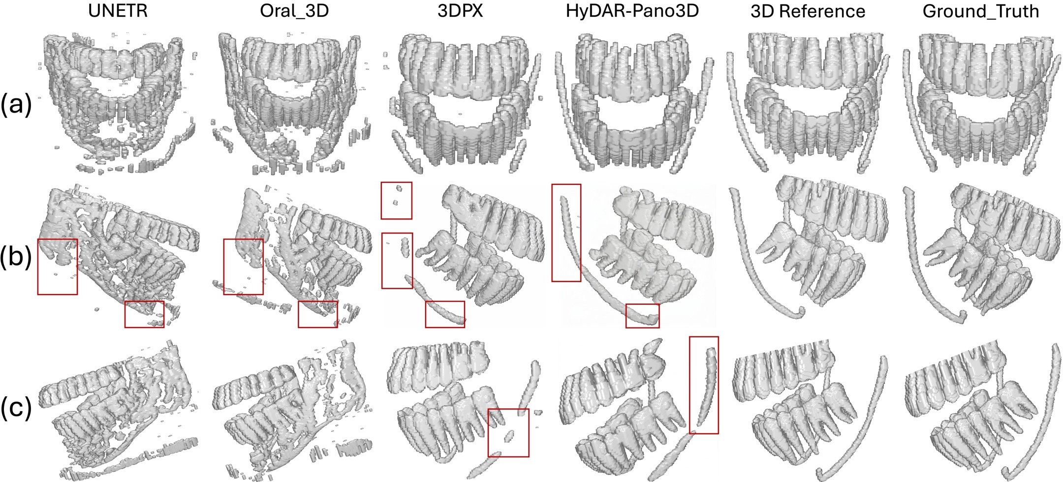
Figure 3: Downstream tooth and IAC segmentation performance—HyDAR-Pano3D reconstructions show increased geometric fidelity and reduced topological errors.
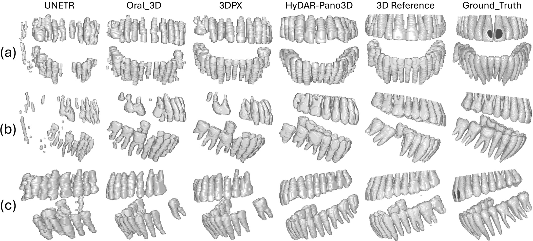
Figure 4: Tooth segmentation on TF dataset; improved recovery of molar and root details seen with HyDAR-Pano3D.
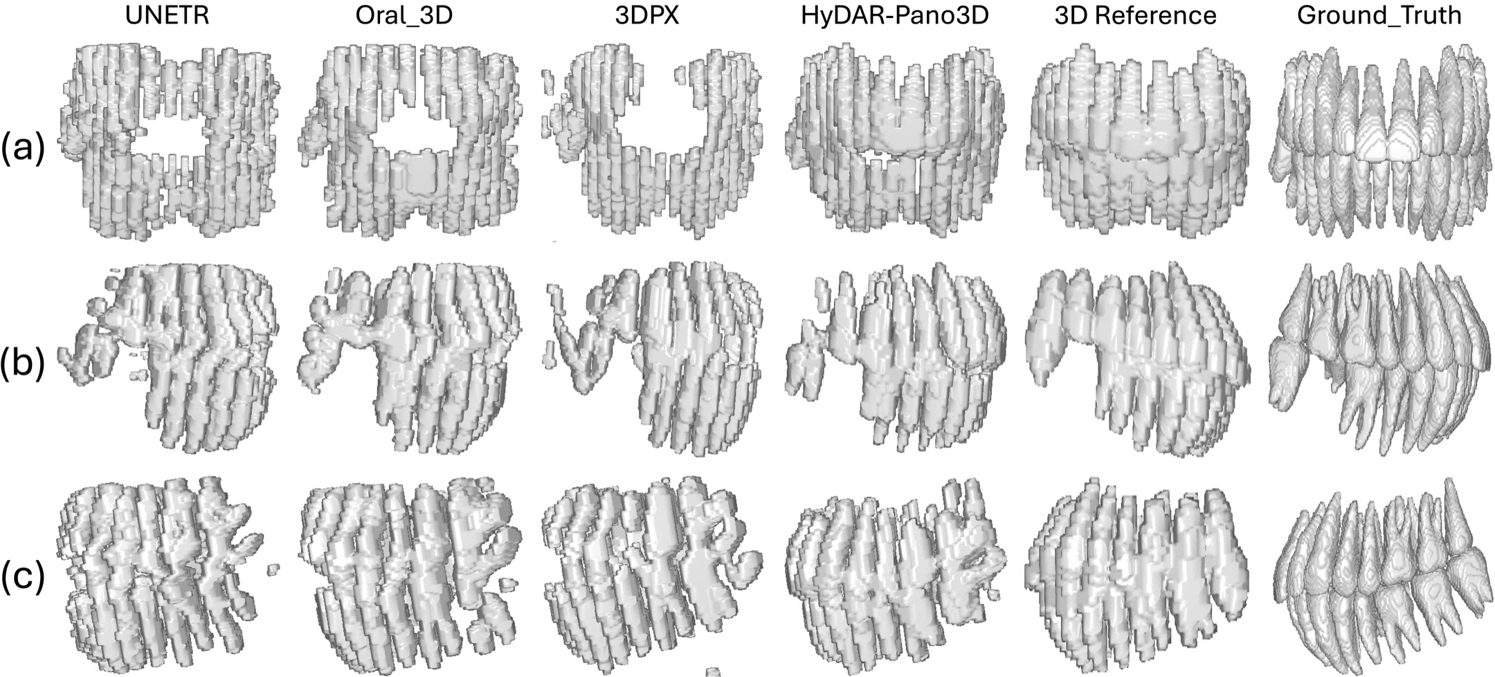
Figure 5: NC dataset segmentation—consistent improvements persist across anatomical and acquisition heterogeneity.
Ablation Analyses and Architectural Insights
Ablation studies establish the criticality of both semantic priors and dual-encoder fusion.
- Substitute foundation models (SAM-ViT-B, MedSAM-lite) yield incremental improvements; however, domain-adapted SemiT-SAM is consistently superior.
- ACF outperforms naive concatenation for feature fusion, particularly in structurally complex regions.
- Dual-encoder models surpass either stream in isolation, confirming the complementary value of local radiographic and global semantic features.
Statistical analysis reveals improvements are robust and significant (paired t-test, p<0.05), and performance gains persist even when canonical to native-space mapping is disabled, confirming the primary contributions originate from the disentangled volumetric inference.
Implications, Limitations, and Future Directions
Pragmatically, HyDAR-Pano3D enables clinically actionable 3D volume synthesis from single PRs, allowing for segmentation-driven diagnostic and planning tasks on populations where CBCT is contraindicated or unavailable. The explicit separation of volumetric appearance and geometric variability represents a strong architectural prior, potentially generalizable to other ill-posed cross-modality inverse problems in medical imaging.
Limitations remain:
- Supervised training leverages synthetic (not clinical) panoramic images, necessitating improved simulation and domain adaptation for deployment.
- Manual dental arch annotation is required for ground truth and could benefit from automation.
- Restoration is regularized for smooth, plausible transformations; extreme pathologies may challenge this approach.
Future research should explore self-supervised or uncertainty-aware formulations, automated arch estimation, stronger domain adaptation, and full integration with clinical decision-support systems.
Conclusion
HyDAR-Pano3D introduces a rigorous disentangled reconstruction framework, combining the strength of vision foundation models for precision anatomy inference with deformation-based restoration for individualized geometry. Empirical evidence across datasets and tasks demonstrates that this bifurcated approach delivers state-of-the-art volumetric fidelity and anatomical integrity, facilitating robust and clinically relevant 3D analysis directly from panoramic radiographs.