Functional Connectivity Dynamics show Resting-State Instability and Rightward Parietal Dysfunction in ADHD
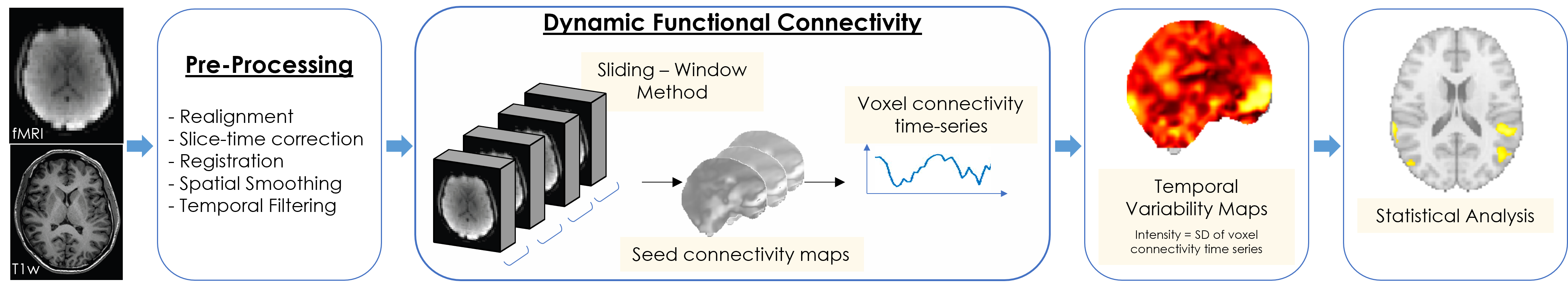
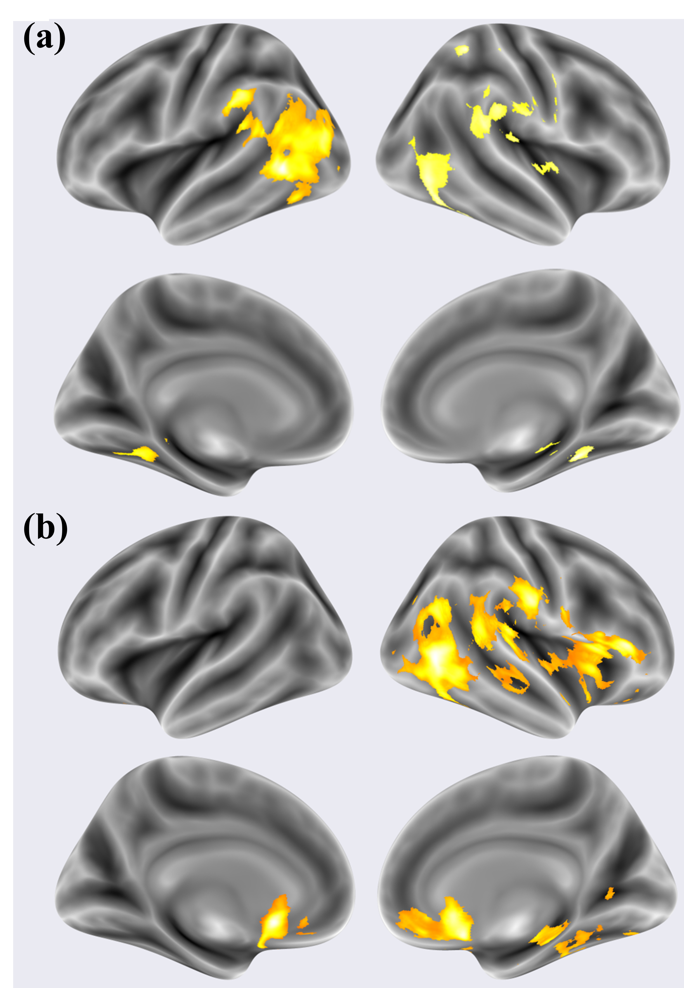
Abstract: Attention Deficit/Hyperactivity Disorder (ADHD) is one of the most common neurodevelopmental disorders in children and is characterised by inattention, impulsiveness and hyperactivity. While several studies have analysed the static functional connectivity in the resting-state functional MRI (rs-fMRI) of ADHD patients, detailed investigations are required to characterize the connectivity dynamics in the brain. In an attempt to establish a link between attention instability and the dynamic properties of Functional Connectivity (FC), we investigated the differences in temporal variability of FC between 40 children with ADHD and 40 Typically Developing (TD) children. Using a sliding-window method to segment the rs-fMRI scans in time, we employed seed-to-voxel correlation analysis for each window to obtain time-evolving seed connectivity maps for seeds placed in the posterior cingulate cortex (PCC) and the medial prefrontal cortex (mPFC). For each subject, the standard deviation of the voxel connectivity time series was used as a measure of the temporal variability of FC. Results showed that ADHD patients exhibited significantly higher variability in dFC than TD children in the cingulo-temporal, cingulo-parietal, fronto-temporal, and fronto-parietal networks ($p_{FWE} < 0.05$). Atypical temporal variability was observed in the left and right temporal gyri, the anterior cingulate cortex, and lateral regions of the right parietal cortex. The observations are consistent with visual attention issues, executive control deficit, and rightward parietal dysfunction reported in ADHD, respectively. These results help in understanding the disorder with a fresh perspective linking behavioural inattention with instability in FC in the brain.
Paper Prompts
Sign up for free to create and run prompts on this paper using GPT-5.
Top Community Prompts
Explain it Like I'm 14
Overview
This paper studies how the brains of children with ADHD connect and “talk” to themselves when they are resting. The authors wanted to see if the way these brain connections change over time is less stable in kids with ADHD compared to kids without ADHD. Their big idea: if attention is shaky in ADHD, maybe the brain’s communication patterns are also more “bouncy” or unstable.
Key Questions
The study asked simple, clear questions:
- Do children with ADHD show more ups and downs in how different brain areas are connected during rest?
- Are certain brain regions—especially those involved in mind-wandering, attention, and self-control—more affected?
- Is there a pattern that matches problems with attention and control often seen in ADHD?
How They Did It (in everyday language)
Think of the brain like a group chat where different teams of friends (brain regions) talk to each other. Even when you’re “resting” and not doing a task, these chats keep going. The researchers looked at how strong these chats were and how much they changed over time.
Here’s the approach, explained simply:
- They used resting-state fMRI, which is like recording a short “movie” of the brain’s activity while the person lies still. The signals come from blood flow (BOLD), which rises and falls as different brain areas are active.
- They focused on two key “hub” areas in the default mode network (DMN), the brain’s mind-wandering network:
- The posterior cingulate cortex (PCC)
- The medial prefrontal cortex (mPFC)
- They measured “functional connectivity,” which means how closely two areas’ activity rises and falls together—like voices in a group chat speaking in sync.
- To study changes over time, they didn’t just look at the whole 6-minute scan at once. Instead, they sliced it into many short time windows (like checking the group chat every few seconds). This is called a sliding-window method.
- In each window, they measured how strongly the seed areas (PCC or mPFC) were connected with every other part of the brain. Then they watched how those connection strengths fluctuated from window to window.
- They used the amount of fluctuation (the standard deviation—think of it as “how jumpy is the signal?”) as a measure of instability or variability in connection over time.
They compared 40 children with ADHD to 40 typically developing children. They used careful statistics that make sure results are very unlikely to be due to chance, and they controlled for age and sex to be fair.
Main Findings and Why They Matter
The key findings are:
- Kids with ADHD showed more variability—more ups and downs—in how their brain areas were connected over time. This happened in networks that link:
- Cingulate regions (involved in monitoring and control) with temporal areas (involved in processing sights, sounds, and meaning)
- Frontal areas (involved in planning and decision-making) with temporal and parietal areas (involved in attention and spatial skills)
- Specific places with more instability included:
- The left and right temporal gyri (important for language, recognizing things, and social/emotional understanding)
- The anterior cingulate cortex (ACC), a key part of the brain’s “executive control” system that helps you focus and manage tasks
- Lateral parts of the right parietal cortex (important for attention, especially shifting and focusing attention in space)
- No areas showed lower variability in ADHD compared to the control group—meaning the pattern was consistently “more unstable” in ADHD.
Why this matters:
- The results match what we know about ADHD: difficulties with attention, staying on task, and self-control.
- The “rightward parietal dysfunction” (more problems on the right side of the parietal lobe) fits with previous brain studies and could be a sign or biomarker to look for in ADHD.
- Overall, the findings offer a brain-based explanation for the “inattention” in ADHD: if the brain’s networks are more unstable, it’s harder to keep attention steady.
Implications and Impact
This research suggests a simple but powerful idea: attention problems in ADHD may come from unstable brain connections during rest, especially in networks that handle mind-wandering, attention, and control. Understanding this could help in several ways:
- Better tools for diagnosis: measuring how “bouncy” brain connections are might help identify ADHD more reliably.
- Targeted treatments: therapy, training, or neurofeedback could aim to stabilize these specific networks.
- More precise research: future studies can focus on the right parietal and ACC/DMN regions to track changes over development or with treatment.
In short, the paper links everyday ADHD symptoms—like trouble focusing—to a clear pattern in the brain: the “group chat” among key areas is more unstable, especially in attention and control networks.
Collections
Sign up for free to add this paper to one or more collections.