- The paper presents an automated method for assessing prostate cancer aggressiveness using multiparametric MRI and ElasticNet regression.
- It extracts 1464 quantitative features from MR images, achieving 58% sensitivity, 87% specificity, and an AUC of 0.73 for aggressive cancer detection.
- The approach bypasses traditional image registration, highlighting DWI lesion boundary roughness as a key predictor for improved diagnostics.
Automated Multiparametric MR Image Analysis for Assessing Prostate Cancer Aggressiveness
Introduction
Prostate cancer (CaP) is a major health concern, being the second leading cause of cancer-related deaths among men in the United States. Current diagnostic techniques, such as prostate-specific antigen (PSA) screening and 'blind' biopsy, have limitations, particularly in differentiating aggressive from non-aggressive cancer. Multiparametric MRI offers a promising avenue for distinguishing aggressive cancers, but is hindered by variability among radiologists and the computational challenges of analyzing high-dimensional imaging data.
Methodology
This research introduces an advanced automated method for assessing the aggressiveness of prostate cancer using multiparametric MRI. Focused on identifying aggressive CaP (Gleason score ≥ 7), the study analyzes quantitative imaging features extracted from T2-weighted MR images, diffusion-weighted images (DWI), and apparent diffusion coefficient maps (ADC).
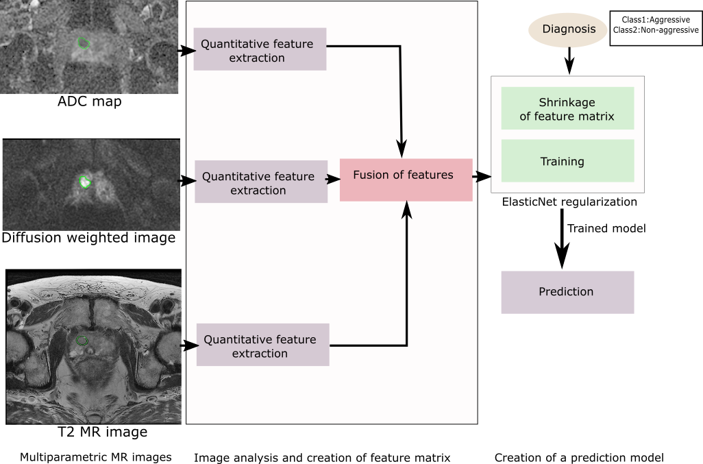
Figure 1: Our prediction model with multiparametric MR image processing pipeline
The methodology involves the creation of a feature matrix comprising 1464 quantitative features, extracted from multiparametric MRI sequences. Post normalization, ElasticNet regularized regression is employed, leveraging both L1 and L2 penalties to preprocess the data and select discriminative features for training the prediction model. This approach circumvents the challenges posed by high-dimensional data and limited sample sizes.
Experimental Results
The study used an MRI dataset from 79 patients, segregated into 40 with aggressive cancer and 39 with less aggressive tumors. The ElasticNet model demonstrated a sensitivity of 58% and a specificity of 87% at the optimum ROC operating point, achieving an AUC of 0.73, which is comparable to methods utilizing elastic registration of MR images. The model identified 44 discriminative features critical for differentiation, with the DWI lesion boundary roughness emerging as the most informative predictor.
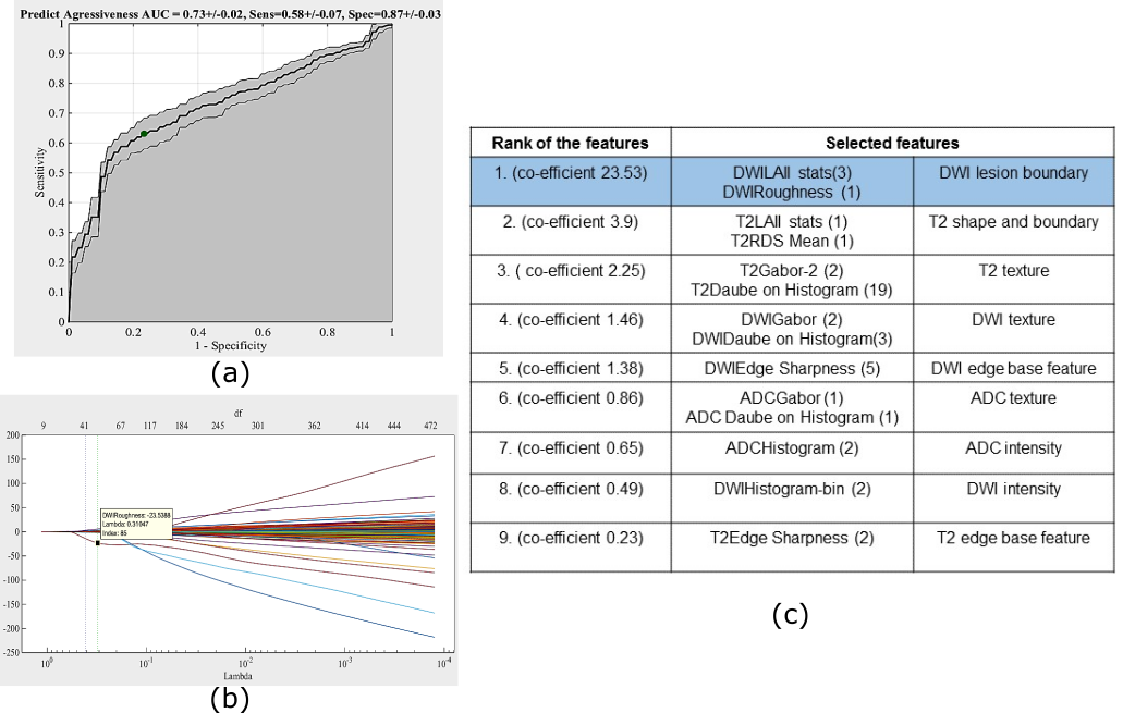
Figure 2: (a) ROC curve, (b) trace plot of coefficient fit, (c) feature ranking
Discussion
This work proposes that real-time feature extraction and automatic classification using multiparametric MRI are feasible without the need for image registration, offering significant improvements over traditional methods. The findings illustrate that DWI is highly informative for assessing CaP aggressiveness. The study highlights potential areas for further exploration, including the evaluation of volumetric data and validation across larger datasets, which may refine the predictive accuracy and broaden the model's applicability.
Conclusion
The introduction of a computerized multiparametric MR image analysis model presents an efficient method for distinguishing between aggressive and non-aggressive prostate cancer. By harnessing sophisticated feature extraction techniques and leveraging advanced regression models, the research paves the way for more accurate diagnostics, potentially informing treatment decisions and improving patient outcomes. Continued exploration and validation could further augment the utility of this approach in clinical settings.