Reduced Gibbs free energy supply hinders brain information processing during mental fatigue
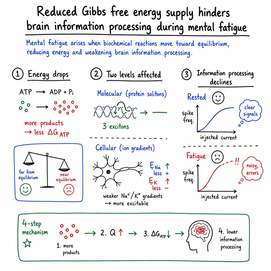
Abstract: Background: Brain information processing deteriorates as cortical neurons gradually transition from a rested state into fatigue. Subjectively, fatigue is experienced as a state of weariness, tiredness, or lack of energy that reduces the ability to work safely and effectively. Objective: In this theoretical paper, we pinpoint the physical origin of brain fatigue in the gradual deterioration of biochemical reaction quotients and transmembrane ion concentration gradients, which increase neuronal excitability and decrease the signal-to-noise ratio in the brain cortex. Methods: Brain performance in a rested state versus fatigue is examined by well-established, data-driven computer models for energy transport inside protein $α$-helices in the presence of thermal noise for different ATP energy states or for pyramidal neuron firing of action potentials under electric stimulation in a rested membrane state versus fatigue. Results: We found that reduced Gibbs free energy supply from ATP hydrolysis impairs the cooperative effect between amide I excitons propagating inside protein $α$-helices, with resulting decreased thermal stability of molecular solitons. Concurrent changes in Nernst reversal potentials for Na+ or K+ ions further led to neuronal hyperexcitability and a higher risk of neuronal depolarization block during mental fatigue. Conclusions: Detailed computational modeling showed that inefficient protein function due to diminished ATP energy status, alters the electrophysiological properties of individual neurons, thereby impairing their information processing capacity for the proper execution of cognitive tasks. Scheduling practices aimed at intermittent recovery of the rested brain state during intellectually challenging work could protect physical and mental wellbeing, prevent burnout, and enhance long-term productivity.
Paper Prompts
Sign up for free to create and run prompts on this paper.
Top Community Prompts
Explain it Like I'm 14
论文概述
这篇论文研究了一个常见的问题:为什么长时间思考、学习或工作后,大脑会变得疲劳,注意力下降,甚至更容易犯错?
作者提出,精神疲劳不仅是“感觉累”,还可能有一个可以用物理学和生物化学解释的原因:脑细胞获得和使用能量的能力逐渐下降,导致神经元传递信息的效率变差。
需要注意的是,这是一篇理论研究。作者主要通过数学计算和计算机模拟来研究疲劳,而不是直接在人体上进行实验。
研究想回答什么问题?
作者主要想了解以下几个问题:
- 大脑疲劳时,细胞能从 ATP 中获得的有效能量是否会减少?
- 能量减少会不会影响神经元内部蛋白质的正常工作?
- 能量不足会怎样改变神经元发放电信号的能力?
- 这些变化是否能够解释注意力下降、反应变慢和认知错误增加?
- 如果经常安排短暂休息,是否可能帮助大脑恢复?
这里的 ATP 可以理解为细胞里的“能量分子”。它有点像电池,但并不是简单地“有多少 ATP 就有多少能量”。ATP 真正能提供多少能量,还取决于细胞中 ATP、ADP 和磷酸盐等物质的比例。
研究方法:作者是怎样研究的?
作者没有直接观察正在考试或工作的人的大脑,而是建立了两个主要的计算模型。
1. 计算 ATP 能提供多少有效能量
作者使用了热力学中的 吉布斯自由能。它表示一个化学反应能够拿出多少能量去完成有用的工作。
可以把它想象成水坝中的水:
- 水位差很大时,水可以推动发电机;
- 水位差变小时,能产生的电就会减少。
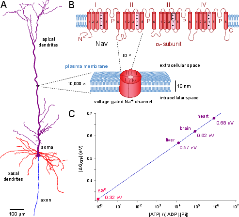
类似地,细胞内 ATP、ADP 和磷酸盐的比例会影响 ATP 分解时能释放多少“可用能量”。作者比较了肝脏、大脑和心脏中的这种能量差异。
2. 模拟能量在蛋白质中的传递
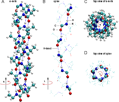
神经元中的许多蛋白质需要能量才能工作。作者使用了一种叫作 Davydov 模型的计算方法,模拟能量如何沿着蛋白质中的螺旋结构传播。
这种能量传播可以想象成体育场里连续移动的“人浪”:
- 如果能量足够,人浪可以稳定地向前移动;
- 如果能量不足,周围的热运动会把它打散。
这种沿蛋白质传播的稳定能量波被称为分子孤子。作者模拟了能量较充足和较不足时,这种波能持续多久、能走多远。
3. 模拟神经元如何发放电信号
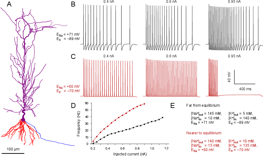
作者还建立了一个完整的海马锥体神经元计算模型。神经元可以通过让带电的钠离子和钾离子进出细胞来产生电信号。
可以把神经元想象成一个有许多自动门的房间:
- 钠离子通道打开时,钠离子进入,帮助神经元“点火”;
- 钾离子通道打开时,钾离子流出,帮助神经元恢复;
- ATP 驱动的钠钾泵则像“清洁和维护工”,把离子重新放回合适的位置。
作者分别模拟了:
- 休息状态:钠离子和钾离子的浓度差较大;
- 疲劳状态:这种浓度差变小,代表离子泵没有完全恢复原来的状态。
作者随后比较了两种情况下神经元的放电频率和稳定性。
主要发现
ATP 能量减少会影响蛋白质中的能量传递
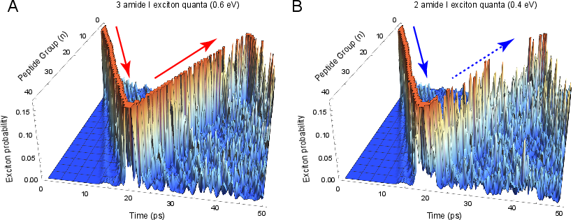
在模拟中,大脑处于较好的能量状态时,ATP 水解大约可以提供 0.62 eV 的能量。这个能量足以帮助形成由三个能量单位组成的稳定分子孤子。
结果显示:
- 三个能量单位组成的孤子可以持续至少约
50皮秒,并传播约45纳米; - 两个能量单位组成的孤子只能持续约
20皮秒,传播距离不到19纳米。
虽然这些距离和时间非常微小,但蛋白质本身也非常小。作者认为,这说明能量不足时,蛋白质可能无法把能量准确地送到需要它的地方,从而降低工作效率。
换句话说,疲劳可能会让神经元里的“微型能量传送带”变得不稳定。
离子浓度差变小会使神经元过度兴奋
在正常休息状态下,细胞内外的钠离子和钾离子浓度差很大。这种差异为神经信号提供了动力。
长时间活动后,如果 ATP 不足,钠钾泵可能无法及时恢复离子分布。作者模拟了这种情况,发现:
- 钠离子和钾离子的“反转电位”发生变化;
- 神经元变得更容易被激活;
- 神经元对外界刺激的反应可能变得不稳定;
- 在刺激太强时,神经元可能进入一种叫作去极化阻滞的状态。
去极化阻滞可以理解为:神经元的“开关”一直处于不正常的开启状态,结果反而无法继续准确地发出新的信号。
大脑的信噪比可能下降
信噪比是指有用信号与杂乱干扰之间的比例。
例如,在教室里听老师讲话时:
- 老师的声音是有用信号;
- 同学说话、空调声和其他杂音是噪声。
如果大脑中的神经元过度兴奋,神经元可能对无关的刺激也作出反应。这样一来,大脑就更难集中注意力、过滤干扰和正确完成任务。
因此,疲劳时可能出现:
- 反应速度变慢;
- 注意力下降;
- 更容易分心;
- 处理信息不够准确;
- 在自己还没有明显感觉疲倦时就已经开始犯小错误。
这些结果为什么重要?
这项研究把“精神疲劳”解释成一个逐渐发展的生理过程,而不是简单的“意志力不够”。
作者提出的过程可以概括为:
- 长时间工作会使 ATP 分解产生的物质逐渐积累;
- ATP 能释放的有效能量减少;
- 蛋白质传递和使用能量的能力下降;
- 离子泵难以维持神经元内外的正常离子差;
- 神经元变得过度兴奋,信息中的噪声增加;
- 大脑处理信息和完成任务的能力下降。
作者还强调,疲劳不是突然从“完全正常”变成“完全不能工作”。它更像一条连续的坡:
- 一开始,变化可能很轻微,自己感觉不到;
- 随着时间增加,错误和分心逐渐变多;
- 如果继续消耗,神经元的工作会受到更严重的影响。
研究的局限
这篇论文主要依靠计算机模型,因此它说明的是一种可能的机制,而不是已经完全证明的人体过程。
例如:
- 模型模拟了单个神经元和蛋白质,但真实大脑有数百亿个相互连接的神经元;
- 论文没有直接测量人在学习或工作时的 ATP 能量变化;
- 真实的疲劳还受到睡眠、压力、情绪、激素和脑区之间合作等因素影响;
- 模型中的某些疲劳状态是根据已有数据推算出来的。
因此,未来还需要人体实验和脑成像研究来检验这些预测。
可能的影响和实际意义
这项研究支持一个简单而实用的建议:在长时间进行高强度脑力活动时,短暂而规律的休息可能比一直坚持到非常疲惫再休息更有帮助。
休息、睡眠、适当运动和减轻压力可能帮助身体:
- 补充和重新分配能量;
- 恢复神经元内外的离子平衡;
- 降低神经元过度兴奋;
- 提高注意力和工作准确性;
- 减少长期压力和职业倦怠。
这对学生、程序员、医生、飞行员和需要持续集中注意力的人都可能有意义。特别是在飞行驾驶或交通控制等工作中,即使一个人主观上还不觉得累,轻微的注意力下降也可能带来严重后果。
总的来说,这篇论文认为:精神疲劳可能是大脑能量供应逐渐下降后,蛋白质和神经元工作效率变差的结果。规律休息并不是浪费时间,而可能是帮助大脑保持准确、高效工作的一种方式。
Knowledge Gaps
Knowledge gaps, limitations, and open questions
The paper leaves the following issues unresolved:
- No direct experimental validation in humans: The proposed link between reduced ATP free energy, altered neuronal excitability, and mental fatigue is not tested using simultaneous measurements of cognitive performance, brain metabolites, ionic gradients, and neuronal activity in human participants.
- ATP concentrations are not measured during mental fatigue: The study assumes that mental work reduces the ATP/ADP/Pi reaction quotient, but does not establish the magnitude, timing, or reversibility of these changes in the human brain during realistic cognitive tasks.
- Use of static metabolite values: Gibbs free energy is calculated from single literature-derived concentrations for whole organs, although ATP, ADP, and Pi vary across brain regions, cell types, subcellular compartments, activity states, and time scales.
- Thermodynamic activities are approximated by concentrations: The calculations do not appear to account for activity coefficients, cytosolic crowding, pH, magnesium binding, temperature variation, or compartment-specific biochemical conditions, all of which can substantially affect the phosphorylation potential.
- Unclear relationship between ATP hydrolysis and protein soliton formation: The paper assumes that ATP free energy is transferred into discrete amide I exciton quanta, but does not demonstrate the biochemical pathway, coupling efficiency, or molecular structure that would connect ATP hydrolysis to Davydov soliton generation in neuronal proteins.
- Davydov’s model has limited biological scope: Simulations use an idealized 40-residue poly-alanine-like -helix with fixed parameters rather than experimentally characterized sequences and structures from neuronal ATPases, ion channels, receptors, cytoskeletal proteins, or synaptic proteins.
- Protein environmental complexity is omitted: The soliton model does not incorporate membrane interfaces, water structuring, lipid interactions, protein tertiary structure, post-translational modifications, heterogeneous damping, local electric fields, or interactions with neighboring proteins.
- Limited stochastic sampling and uncertainty analysis: The reported soliton lifetimes are based on a small number of simulations using shared Wiener processes, without confidence intervals, parameter sweeps, convergence tests, or sensitivity analysis across thermal-noise realizations.
- Questionable discrete energy threshold: The conclusion that a decrease below necessarily reduces excitation from three to two amide I quanta assumes strictly quantized, directly allocated ATP energy and does not examine partial energy transfer, multiple ATP molecules, nonlinear coupling, or alternative dissipation pathways.
- Soliton lifetimes are not experimentally confirmed: The predicted 20–50 ps lifetimes and 19–45 nm transport distances have not been compared with direct measurements of vibrational energy transport in relevant neuronal proteins under physiological conditions.
- Ion-gradient depletion is imposed rather than modeled dynamically: The neuronal simulations prescribe fatigue-state reversal potentials but do not simulate ATP production, pump kinetics, ion-channel activity, sodium and potassium accumulation, diffusion, buffering, or extracellular-space dynamics over time.
- The selected “fatigue” reversal potentials lack empirical justification: The values mV and mV are based on a hypothetical exchange of 5 mM intracellular potassium for sodium, but the paper does not show that these concentration changes occur in healthy humans during mental fatigue.
- Other relevant ions and transporters are incompletely integrated: The analysis emphasizes sodium and potassium while not dynamically linking calcium gradients, chloride, bicarbonate, proton gradients, glutamate transport, calcium pumps, exchangers, and intracellular buffering to the proposed fatigue mechanism.
- Single-neuron generalizability is unknown: Results from one morphologically reconstructed CA1 pyramidal neuron cannot establish effects across cortical pyramidal neurons, interneurons, hippocampal cells, thalamic neurons, or other neuronal classes with different channel distributions and energy demands.
- Synaptic computation is not modeled: The study examines somatic current injection and firing frequency but does not test how fatigue-related changes affect synaptic integration, dendritic computation, plasticity, inhibition–excitation balance, spike timing, network oscillations, or sensory coding.
- Signal-to-noise reduction is asserted but not quantified: The paper interprets increased excitability as a lower signal-to-noise ratio without defining a noise model or measuring information-theoretic quantities such as mutual information, coding accuracy, variability, or error rates.
- No network-level demonstration of cognitive impairment: The proposed cellular changes are not propagated into network or whole-brain models that could predict effects on attention, working memory, decision-making, reaction time, or task accuracy.
- No behavioral dose–response relationship: The paper does not determine how specific changes in ATP free energy or reversal potentials translate into measurable cognitive-performance declines or identify thresholds for clinically meaningful impairment.
- Alternative mechanisms of mental fatigue are not systematically compared: Sleep pressure, neuromodulators, inflammation, stress hormones, motivation, vascular regulation, astrocytic metabolism, neurotransmitter depletion, and psychological effort are discussed only briefly and are not incorporated into a competing or integrated model.
- Causal direction remains unresolved: It is unclear whether metabolic and ionic changes initiate mental fatigue, result from sustained neural activity, or interact bidirectionally with cognitive and motivational processes.
- Spatial propagation from local to global fatigue is unspecified: The paper acknowledges subcellular compartmentalization but does not model how local ATP depletion and ionic disturbances spread across synapses, dendrites, cortical columns, brain regions, or distributed cognitive networks.
- Temporal dynamics and recovery are not characterized: The simulations do not establish how quickly ATP pools, ionic gradients, protein function, and cognitive performance deteriorate or recover during breaks, naps, sleep, or repeated work–rest cycles.
- The proposed rest-scheduling intervention is untested: The recommendation for shorter and more frequent breaks is theoretical; optimal break duration, frequency, timing, task dependence, and individual variability remain unknown.
- No biomarkers for early “subtle incapacitation” are validated: The paper proposes that impairment may precede detectable EEG or fMRI changes but does not identify or test a sensitive early marker, such as spectroscopy, metabolic imaging, high-density electrophysiology, pupillometry, or behavioral variability.
- EEG and fMRI inferences are overstated without empirical coupling: The manuscript does not establish a quantitative mapping between the modeled ionic or metabolic changes and EEG, BOLD, or magnetic-resonance spectroscopy signals.
- Healthy fatigue is not differentiated from pathological fatigue empirically: Although neuropsychiatric and neurological diseases are excluded conceptually, the model does not test whether its predictions distinguish reversible mental fatigue from depression, anxiety, multiple sclerosis, Parkinson’s disease, or other disorders.
- Sex, age, fitness, sleep history, nutrition, and metabolic health are not considered: These factors may alter ATP availability, mitochondrial reserve, ion-pump capacity, fatigue susceptibility, and recovery, but are absent from the model.
- Metabolic supply and blood flow are simplified: Glucose delivery, oxygen availability, lactate metabolism, mitochondrial respiration, cerebral blood flow, and neurovascular coupling are not modeled, limiting interpretation of ATP depletion as the primary energy constraint.
- No comparison with experimental fatigue paradigms: The proposed mechanism is not evaluated across different cognitive tasks, workload intensities, durations, sensory demands, or forms of fatigue, so its domain of applicability is unclear.
- Model parameter uncertainty is not propagated to conclusions: The robustness of the predicted transition to hyperexcitability or depolarization block under uncertainty in channel densities, temperature, membrane properties, pump rates, and metabolite concentrations remains undetermined.
- The depolarization-block prediction lacks physiological corroboration: The paper does not show whether healthy mental fatigue reaches the modeled ionic state or produces depolarization block in vivo, as opposed to more modest changes in firing reliability or network coordination.
- Energy accounting is incomplete: The analysis does not quantify the fraction of total neuronal ATP consumption attributable to the modeled proteins, ion pumps, synaptic transmission, calcium handling, glial support, and maintenance processes.
- The proposed mechanism is not shown to be necessary or sufficient: It remains unresolved whether restoring ATP availability or ionic gradients alone would prevent fatigue, and whether these changes are sufficient to reproduce fatigue-related cognitive deficits in the absence of other mechanisms.
Practical Applications
Immediate Applications
The paper is theoretical and does not validate a clinical diagnostic or intervention protocol. Nevertheless, its central implication—that cognitive performance may deteriorate before a person consciously notices fatigue—supports several low-risk applications based on existing practices and technologies.
- Workplace scheduling for cognitively demanding tasks — occupational health, education, software, and professional services
- Implement short, frequent recovery periods during sustained mental work rather than relying only on long, infrequent breaks.
- Apply this to programming, examination periods, legal or financial analysis, scientific research, call centers, and administrative work.
- Workflows could use scheduled intervals—for example, alternating focused work with brief rest, movement, hydration, or low-demand activities—especially during tasks requiring sustained attention.
- Category rationale: Immediate Application, because scheduling and break-management interventions require no new biomedical technology.
- Dependencies and assumptions: The paper does not establish an optimal break duration or frequency. Effectiveness may vary with sleep, stress, workload, individual metabolism, task type, and the distinction between physical and mental fatigue.
- Fatigue-risk management in safety-critical occupations — aviation, air-traffic control, transportation, healthcare, and industrial operations
- Incorporate mandatory cognitive-rest intervals, shift-design rules, and limits on uninterrupted high-attention work.
- Combine existing self-report tools, reaction-time tests, vigilance tasks, and workload monitoring with conservative scheduling policies.
- Operational protocols could require task rotation or independent verification of decisions after prolonged periods of concentration.
- Category rationale: Immediate Application, because fatigue-risk-management systems and procedural safeguards already exist and can be strengthened without waiting for a validated biomarker.
- Dependencies and assumptions: The paper’s proposed early “subtle incapacitation” is not directly measured in humans. Any operational threshold would require empirical validation to avoid excessive interruptions or false alarms.
- Educational study and examination design — schools, universities, and professional training
- Schedule demanding examinations, laboratory sessions, and high-stakes simulations with planned recovery periods.
- Use shorter learning blocks, alternating subjects or cognitive modalities, and deliberate pauses before complex reasoning or memory-retrieval tasks.
- Learning-management systems could remind students to pause after prolonged periods of continuous engagement.
- Category rationale: Immediate Application, because these are behavioral and instructional-design changes consistent with the paper’s prophylactic recommendation.
- Dependencies and assumptions: The model concerns neuronal energy and excitability, not specific learning outcomes. Educational benefits should therefore be evaluated using accuracy, retention, and response-time measures rather than presumed from the theory alone.
- Personal digital fatigue-management tools — consumer software and daily life
- Develop calendar, browser, or desktop tools that detect extended uninterrupted screen work and recommend brief recovery periods.
- Features could include task-aware reminders, reduced notification density, visual rest periods, brief movement prompts, and end-of-day workload summaries.
- Such tools should treat subjective alertness as insufficient by also encouraging objective checks such as reaction-time or attention tests.
- Category rationale: Immediate Application, because the software can be implemented with existing devices and does not need to estimate ATP or ion concentrations directly.
- Dependencies and assumptions: Screen time is only a proxy for cognitive load. Privacy, user adherence, accessibility, and the risk of excessive or poorly timed reminders must be considered.
- Use of existing fatigue measurements in research and occupational screening — academia and applied neuroscience
- Pair cognitive-performance tests with EEG, reaction-time measures, eye tracking, heart-rate variability, or magnetic-resonance spectroscopy in controlled studies.
- The paper suggests that EEG and macroscopic metabolic measures may detect fatigue relatively late; therefore, researchers can compare them with earlier behavioral changes to identify more sensitive indicators.
- Existing NEURON and ModelDB resources can be used to reproduce or extend the paper’s neuron simulations under different ionic conditions.
- Category rationale: Immediate Application, because the models and measurement technologies already exist.
- Dependencies and assumptions: Computational neuronal behavior does not automatically map onto whole-person cognition. Human validation, standardized fatigue paradigms, and control of sleep, nutrition, medication, stress, and disease status are necessary.
- Model-based interpretation of neuronal energy constraints — computational neuroscience and biomedical education
- Use the CA1 pyramidal-neuron model to teach how ATP-dependent ion pumping, Nernst potentials, firing frequency, and depolarization block are related.
- The model can support hypothesis generation for experiments on repetitive stimulation, ionic-gradient recovery, and neuronal hyperexcitability.
- Category rationale: Immediate Application, because the relevant simulation environment and model parameters are publicly available.
- Dependencies and assumptions: The simulations use a specific neuron morphology and selected reversal-potential conditions. They should not be treated as a general model of all cortical neurons or as a clinical predictor.
- Conservative public-health guidance on recovery behaviors — policy and daily life
- Promote regular sleep, manageable workloads, stress reduction, physical activity, and brief recovery periods as general fatigue-prevention measures.
- Employers and public institutions can frame these practices as performance and safety measures rather than as responses only after subjective exhaustion appears.
- Category rationale: Immediate Application, because these recommendations are low-risk and align with established fatigue-management practice.
- Dependencies and assumptions: The paper does not show that a particular diet, supplement, nap duration, or lifestyle intervention restores neuronal Gibbs free energy. Claims about ATP enhancement or cognitive improvement would require separate clinical evidence.
Long-Term Applications
The following applications require validation in humans, improved models, or development of reliable noninvasive measurements. The paper’s computational findings should be regarded as mechanistic hypotheses rather than established diagnostic thresholds.
- Early-warning systems for cognitive incapacitation — aviation, transport, medicine, defense, and industrial safety
- Develop multimodal systems that combine reaction-time variability, error patterns, EEG features, eye movements, speech or typing behavior, and workload history.
- A future system could estimate a person’s probability of reduced signal-to-noise processing before obvious sleepiness or self-reported fatigue, then recommend a break or transfer responsibility.
- Potential products include cockpit fatigue monitors, clinician-alert systems, driver-assistance interfaces, and operator “fit-for-task” dashboards.
- Category rationale: Long-Term Application, because the paper predicts early cellular changes but does not provide a validated human biomarker or decision threshold.
- Dependencies and assumptions: Reliable prediction would require demonstrating that the proposed ionic and metabolic mechanism produces reproducible macroscopic signatures and that these signatures predict real-world errors.
- Personalized computational models of brain-energy status — digital health and precision medicine
- Construct individual models linking workload, sleep, nutrition, metabolic measurements, EEG, and cognitive performance to estimated ATP reaction quotients and ionic-gradient recovery.
- Such models might generate personalized recommendations for work-rest timing or identify unusually slow recovery.
- Category rationale: Long-Term Application, because intracellular ATP, ADP, phosphate, and ionic concentrations are compartmentalized and cannot currently be inferred accurately in routine settings.
- Dependencies and assumptions: The model would need validated relationships between peripheral or imaging measurements and local neuronal energy states. Individual differences in mitochondrial function, vascular supply, medications, and disease would be important confounders.
- Noninvasive fatigue biomarkers based on metabolic and electrophysiological measurements — healthcare and neuroscience
- Investigate whether combinations of EEG, functional MRI, magnetic-resonance spectroscopy, diffusion or metabolic imaging, and behavioral testing can detect the progression described in the paper.
- Candidate markers could include altered glutamate/glutamine or creatine-phosphate signals, changes in network synchronization, and electrophysiological signatures associated with hyperexcitability.
- Category rationale: Long-Term Application, because current techniques have limited spatial, temporal, or biochemical specificity and may detect fatigue only after substantial changes have occurred.
- Dependencies and assumptions: A biomarker must distinguish reversible mental fatigue from sleep deprivation, depression, anxiety, neurological disease, medication effects, and structural neuronal injury.
- Therapeutic strategies targeting neuronal energy and ionic homeostasis — clinical neuroscience and pharmacology
- The mechanism suggests possible research directions involving mitochondrial support, creatine-phosphate buffering, ion-pump function, glutamate clearance, or modulation of pathological hyperexcitability.
- Potential interventions could include pharmacological agents, metabolic therapies, neuromodulation, or targeted rehabilitation for disorders in which fatigue and neuronal hyperexcitability coexist.
- Category rationale: Long-Term Application, because the paper does not test treatments and does not establish that increasing ATP availability or altering ion gradients would improve cognition safely.
- Dependencies and assumptions: Increasing excitability or metabolic activity indiscriminately could worsen excitotoxicity, seizures, oxidative stress, or disease-related injury. Clinical development would require dose-response studies and safety testing.
- Energy-aware artificial neural networks and neuromorphic hardware — artificial intelligence, robotics, and edge computing
- Translate the paper’s principle of performance degradation under diminishing energy reserves into adaptive systems whose signal processing becomes more conservative as battery or computational energy declines.
- Applications could include robots that reduce task complexity when power is low, autonomous vehicles that request human assistance, and edge-AI devices that lower processing load while preserving safety-critical functions.
- Category rationale: Long-Term Application, because this is an engineering analogy rather than a direct biological application of the reported results.
- Dependencies and assumptions: The analogy between ATP-limited neuronal processing and electronic energy constraints must be formalized. Safety requires predictable graceful degradation and cannot rely on subjective fatigue-like indicators.
- Molecular bioengineering for energy transport in proteins — biotechnology and nanomedicine
- The Davydov-model results could motivate investigation of how protein structure, coupling strength, and local energy supply affect vibrational energy transport.
- Future tools might include simulations or engineered protein scaffolds designed to improve energy transfer over nanometer distances in enzymes, molecular motors, biosensors, or synthetic biological circuits.
- Category rationale: Long-Term Application, because the modeled molecular solitons have not been demonstrated as a controllable, general-purpose energy-transfer mechanism in living human neurons.
- Dependencies and assumptions: The conclusions depend on model parameters, thermal-noise assumptions, and the applicability of the Davydov framework to real heterogeneous proteins. Experimental confirmation at physiological conditions is essential.
- Policy standards based on objective cognitive-fatigue thresholds — labor regulation and public safety
- Regulators could eventually establish evidence-based limits for uninterrupted cognitive work, mandatory recovery intervals, and monitoring requirements in safety-critical jobs.
- Policies might distinguish between subjective tiredness and objectively measurable performance degradation, as emphasized by the paper.
- Category rationale: Long-Term Application, because regulatory thresholds require large longitudinal studies linking physiological markers and fatigue scores to accidents, errors, and productivity.
- Dependencies and assumptions: Standards must account for occupational diversity, disability and privacy rights, measurement error, worker autonomy, and the possibility that monitoring could be used punitively rather than preventively.
- Clinical differentiation of reversible fatigue from neurological or psychiatric illness — healthcare
- A validated thermodynamic and electrophysiological framework could help clinicians distinguish ordinary reversible fatigue from fatigue associated with depression, anxiety, multiple sclerosis, Parkinson’s disease, or other conditions.
- This could support treatment selection and monitoring by combining cognitive testing with metabolic, electrophysiological, and clinical data.
- Category rationale: Long-Term Application, because the paper explicitly excludes irreversible neuronal injury and does not establish diagnostic criteria.
- Dependencies and assumptions: The proposed mechanism may overlap with multiple disease processes. Clinical use would require diagnostic accuracy studies, disease-specific models, and evidence that the distinction changes patient outcomes.
Glossary
- Action potential: A rapid, transient electrical impulse that travels along a neuron’s axon. “the neuron fires an action potential”
- Adenosine diphosphate (ADP): A nucleotide produced when ATP loses one phosphate group. “adenosine diphosphate (ADP) and inorganic phosphate”
- Adenosine triphosphate (ATP): The principal cellular molecule used to transfer chemical free energy. “hydrolysis of adenosine triphosphate (ATP)”
- Amide I exciton: A quantized vibrational excitation associated mainly with peptide carbonyl groups. “individual amide I excitons have an energy of ”
- Amide II band: A vibrational mode of peptide bonds involving primarily N–H bending and C–N stretching. “energy transport in the form of amide I (--CO--) and amide II band (--NH--) vibrations”
- Axonal hillock: The region where a neuron’s axon begins and where action potentials are typically initiated. “the membrane potential in the axonal hillock”
- Basal dendrite: A dendritic branch extending from the lower or basal region of a neuron’s soma. “including soma, apical dendrites, basal dendrites and axon”
- Boltzmann constant: A physical constant relating temperature to energy at the molecular scale. “$k_{\text{B} = 1.380649 \times 10^{-23}~\text{J K}^{-1}$ is the Boltzmann constant”
- Blood oxygenation level-dependent (BOLD) signal: An fMRI signal produced by changes in blood oxygenation associated with neural activity. “the blood oxygenation level-dependent (BOLD) signal measures the brain activity indirectly”
- CA1 pyramidal neuron: A principal excitatory neuron found in the CA1 region of the hippocampus. “A morphologically complete model of CA1 pyramidal neuron was simulated”
- Covalent bond: A chemical bond formed through the sharing of electron pairs between atoms. “the lateral coupling of the lattice of peptide groups due to covalent bonds in the protein backbone”
- Creatine kinase: An enzyme that transfers phosphate groups between creatine phosphate and ADP or ATP. “Local depletion of ATP can be temporarily counteracted by brain creatine kinase”
- Creatine phosphate: A phosphorylated creatine molecule that serves as a short-term cellular energy reserve. “which consumes compartmentalized depots of creatine phosphate”
- Cytosol: The aqueous intracellular fluid surrounding cellular organelles. “taking place inside neuronal cytosol”
- Depolarization block: A state in which sustained membrane depolarization prevents a neuron from generating further action potentials. “a higher risk of neuronal depolarization block during mental fatigue”
- Dipole–dipole coupling: An interaction between oscillating or permanent electric dipoles that enables energy transfer. “ is the nearest neighbor dipole-dipole coupling energy”
- Electroencephalography (EEG): A technique that records electrical activity of the brain using electrodes on the scalp. “Alterations in excitability of individual neurons accumulate in time and can be detected at the macroscopic level using electroencephalography (EEG)”
- Electromotive force: The potential difference that drives an electric current, including across a biological membrane. “electromotive forces for different ion types”
- Excitatory postsynaptic potential (EPSP): A temporary postsynaptic depolarization that increases the probability of neuronal firing. “excitatory postsynaptic potentials (EPSPs)”
- Exciton: A quasiparticle representing a coupled excitation that can transport energy through a material. “the cooperative effect between amide I excitons”
- Excitotoxicity: Cellular injury or death caused by excessive excitatory stimulation, often involving glutamate. “prolonged glutamate release can lead to neuronal hyperexcitation and excitotoxicity”
- Exergonic reaction: A chemical reaction that releases free energy and has a negative Gibbs free-energy change. “Since exergonic reactions release energy into the environment”
- Fluctuation–dissipation theorem: A principle relating random thermal fluctuations in a system to its dissipative processes. “which obey the fluctuation-dissipation theorem”
- f-I curve: A graph relating a neuron’s firing frequency to the injected current. “Comparison of the f-I curves far from equilibrium or nearer to equilibrium”
- Gated ion channel: A membrane protein whose ion permeability changes in response to a stimulus such as voltage. “Most of the ion channels are gated by changes in the transmembrane voltage”
- Gibbs free energy: Thermodynamic energy available from a system to perform useful work at constant temperature and pressure. “The Gibbs free energy equation relates the change in Gibbs free energy”
- Glutamatergic neuron: A neuron that uses glutamate as its primary excitatory neurotransmitter. “with of the cortical neurons being glutamatergic”
- Hyperarousal: A state of excessive physiological or neural activation. “it can lead to a state of hyperarousal”
- Hyperexcitability: An abnormally increased tendency of neurons or other excitable cells to respond to stimulation. “This leads to neuronal hyperexcitability”
- Inorganic phosphate: The phosphate species released during ATP hydrolysis. “adenosine diphosphate (ADP) and inorganic phosphate ($\text{P}_{\text{i}$)”
- Ion concentration gradient: A difference in ion concentration across a membrane that can store electrochemical potential energy. “transmembrane ion concentration gradients”
- Ion channel: A membrane-spanning protein that permits selected ions to cross the cell membrane. “the opening or closing of different types of ion channels”
- Ionic pump: A membrane protein that uses energy, typically from ATP, to transport ions against their concentration gradients. “The individual ionic Nernst reversal potentials are dynamically regulated by ionic pumps”
- Langevin equation: A dynamical equation incorporating both deterministic forces and random thermal fluctuations. “the system of stochastic Langevin equations of motion”
- Molecular soliton: A localized, stable wave of molecular excitation that propagates without rapidly dispersing. “with resulting decreased thermal stability of molecular solitons”
- Molar concentration: The amount of a substance per unit volume of solution, commonly expressed in moles per liter. “the concentrations of all aqueous solutions are 1 molar (M)”
- Molecular pharmacology: The study of how drugs interact with molecules and biochemical processes in living systems. “His experience and research interests encompass topics of molecular neuroscience, molecular pharmacology”
- Morphological compartment: A structurally distinct region of a neuron, such as a soma, dendrite, or axon. “individual compartments such as individual dendritic spines”
- Nernst reversal potential: The membrane voltage at which the electrical and chemical forces on a particular ion balance, producing no net ionic current. “Concurrent changes in Nernst reversal potentials for or ions”
- Neurotransmitter: A chemical messenger released by neurons to communicate with other cells. “Glutamate is the major excitatory neurotransmitter in the neocortex”
- Normoergic compound: A compound whose hydrolysis releases a comparatively ordinary amount of free energy. “ATP hydrolysis has large negative compared to normoergic compounds”
- Phosphorylation potential: The free-energy difference associated with ATP synthesis from ADP and inorganic phosphate. “which determines the phosphorylation potential $\Delta G_{\text{P} = -\Delta G_{\text{ATP}$”
- Pyramidal neuron: A neuron with a pyramid-shaped soma and characteristic apical and basal dendrites, common in the cerebral cortex and hippocampus. “for pyramidal neuron firing action potentials under electric stimulation”
- Reaction quotient: A ratio describing the relative activities or concentrations of products and reactants in a chemical reaction at a given moment. “The Gibbs free energy equation relates the change in Gibbs free energy measured in electronvolts (eV) to the reaction quotient ”
- Signal-to-noise ratio: The strength of a meaningful signal relative to irrelevant fluctuations or background noise. “decrease the signal-to-noise ratio in the brain cortex”
- Soma: The cell body of a neuron, containing its nucleus and most organelles. “In neuronal dendrites and soma”
- Stochastic Wiener process: A continuous-time mathematical model of random motion, commonly used to represent Brownian noise. “ are independent real-valued continuous-time stochastic Wiener processes”
- Synaptic plasticity: The ability of synapses to change their strength in response to activity. “the high levels of ATP required for synaptic plasticity”
- Thermodynamic activity: An effective concentration used to account for nonideal behavior in thermodynamic calculations. “the dimensionless thermodynamic activities of , and $|\text{P}_{\text{i}|$”
- Thermodynamic equilibrium: A state in which no net macroscopic thermodynamic change occurs. “there is no change in Gibbs free energy, ”
- Thermal bath: An environment that exchanges energy with a system and maintains it at a specified temperature. “in the presence of a thermal bath with temperature ”
- Thermal noise: Random fluctuations caused by microscopic thermal motion. “in the presence of thermal noise”
- Transmembrane voltage: The electrical potential difference across a cell membrane. “Most of the ion channels are gated by changes in the transmembrane voltage”
- Voltage-gated ion channel: An ion channel whose opening probability depends on the membrane voltage. “Voltage-gated ion channels”
- Voltage sensor: A charged structural region of an ion channel that detects changes in membrane voltage. “acts as a voltage sensor”
- Voltage-gated sodium channel: A membrane protein that selectively conducts sodium ions in response to changes in membrane voltage. “Voltage-gated ion channels ($\text{Na}_{\text{v}$)”
- Wiener process: A stochastic process characterized by continuous paths and normally distributed independent increments. “the same set of 120 randomly generated Wiener processes ”
- α-helix: A protein secondary structure consisting of a helical arrangement of amino-acid residues stabilized by hydrogen bonds. “energy transport inside protein -helices”
Collections
Sign up for free to add this paper to one or more collections.