- The paper presents STRMSR, a novel cardiac MRI super-resolution method that combines reference-guided matching with memory propagation to enhance volumetric consistency.
- It employs a dual-branch Swin Transformer along with coarse-to-fine contextual matching (CFCM) and patch-wise dynamic feature aggregation (PDFA) to accurately transfer high-frequency anatomical details.
- Experimental results on the WHS dataset show statistically significant PSNR improvements and better preservation of myocardial boundaries compared to existing methods.
Reference-and-Memory-Guided Through-Plane Super-Resolution for Cardiac MRI
Introduction
The paper "Cardiac MRI Through-Plane Super-Resolution Guided by Reference and Memory" (2607.07581) addresses a fundamental challenge in clinical cardiac MRI (CMR): the acquisition constraint that results in high in-plane but coarse through-plane resolution due to breath-hold and cardiac-motion limitations. This anisotropy restricts reliable 3D volumetric analysis and downstream diagnosis. The standard solution involves two sequential steps—slice alignment and through-plane super-resolution (SR)—but existing methods often fail to efficiently leverage auxiliary high-resolution (HR) views or enforce cross-slice consistency, leading to blurred reconstructions and inter-slice discontinuities.
To advance SR in cardiac MRI, the authors propose STRMSR, a reference- and memory-guided SR architecture that incorporates coarse-to-fine contextual matching (CFCM) and patch-wise dynamic feature aggregation (PDFA) to robustly transfer anatomical details between misaligned views. A memory bank stores intermediate SR results, enabling slice-to-slice propagation that improves volumetric coherence. STRMSR is evaluated on the WHS cardiac MRI dataset under two clinically realistic reference protocols, demonstrating superior performance particularly at high upsampling factors.
Methodological Overview
STRMSR employs a dual-branch Swin Transformer encoder that extracts features from both LR target and HR reference images. The reference branch preserves resolution-specific details. Initial LR inputs are center-copy upsampled and encoded at multiple scales.
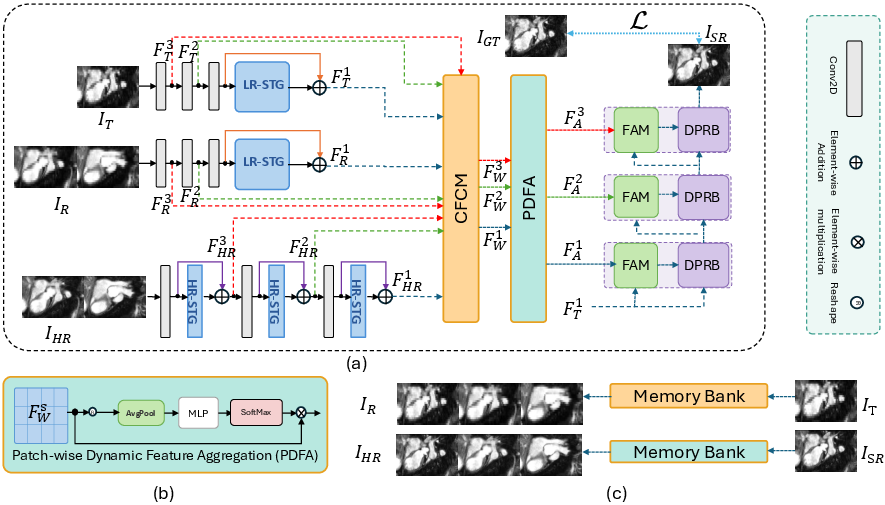
Figure 1: STRMSR architecture: (a) CFCM for robust correspondence, (b) PDFA for patch-wise aggregation, (c) Memory bank storing intermediate SR outputs for propagation.
Coarse-to-Fine Contextual Matching (CFCM)
STRMSR establishes correspondence via multi-dilation normalized correlation at the coarsest level, partitioning features into k×k blocks and refining matches through local dense patch matching. The process iteratively propagates block centers to finer scales, ensuring consistent spatial alignment.
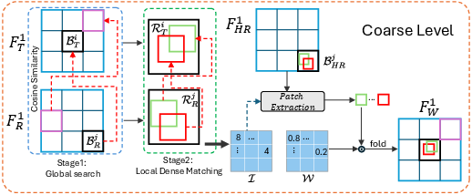
Figure 2: CFCM illustration at the coarsest scale, showing block-wise matching strategy and confidence map construction.
Patch-wise Dynamic Feature Aggregation (PDFA)
PDFA fuses warped reference and memory feature maps using a content-adaptive mixture-of-experts formulation. Local patch descriptors inform a lightweight MLP, which generates softmax-normalized weights for selective feature transfer. This suppresses inconsistent or unreliable matches attributable to anatomical or spatial discrepancies.
Memory-Based SR Propagation
STRMSR treats the LR volume as a temporal sequence. Once the first slice is super-resolved using reference images, its result is deposited into a FIFO memory bank. Subsequent slices benefit from reference-guided aggregation supplemented by memory-based propagation, enforcing inter-slice consistency and enhancing volumetric coherence.
Experimental Validation and Numerical Results
The WHS cardiac MRI dataset is used to benchmark STRMSR under two reference protocols: (i) WHS-Ortho, where dense orthogonal-plane reference volumes provide robust coverage; (ii) WHS-LAX, utilizing sparse long-axis chamber views.
STRMSR consistently outperforms all baselines (Bicubic, MsFF-Net, MINet, MASA, McMRSR), especially at the ×8 upsampling factor where reference cues are sparse and high-frequency detail recovery is most challenging. In WHS-Ortho (×8), STRMSR improves PSNR by +0.97\,dB over McMRSR and +0.55\,dB over MsFF-Net. All improvements at ×8 are statistically significant (p<0.001).
Qualitative evaluations reveal STRMSR’s ability to preserve intricate myocardial boundaries and linear anatomical features that competing methods either blur or omit. Memory-based propagation further yields error maps with lower residuals and temporal profiles evidencing consistent reconstruction across slices.
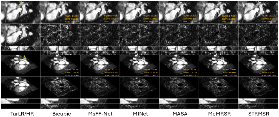
Figure 3: Qualitative comparison on WHS-Ortho and WHS-LAX; SR images, error maps, temporal profiles, and temporal error maps highlight STRMSR's edge sharpness and consistency.
Ablative Analysis
Dropping CFCM in favor of direct LR-level correspondence leads to the greatest PSNR decline, evidencing the necessity of iterative refinement. Removing PDFA reduces robustness in patch selection, while omitting the memory module erodes volumetric consistency. The complementary contributions of CFCM, PDFA, and memory propagation collectively produce the highest SR fidelity.
Implications and Future Directions
The STRMSR framework advances cardiac MRI SR by combining reference-guided matching with memory-based inter-slice propagation, establishing robust volumetric consistency and improved anatomical fidelity even under severe through-plane degradation. Practically, this enables more diagnostic 3D reconstructions from routinely acquired 2D cine stacks. Theoretically, STRMSR provides a generic backbone adaptable to other anisotropic MRI modalities, multi-contrast fusion, or volumetric SR tasks in neuroimaging.
Future research may extend STRMSR to handle more diverse anatomical geometries, integrate unsupervised alignment, or further optimize patch aggregation strategies for computational efficiency. Incorporating spatio-temporal dynamics or multimodal reference data may also enhance SR performance and clinical applicability.
Conclusion
STRMSR sets a new standard for reference- and memory-guided through-plane SR in cardiac MRI, consistently outstripping baselines at clinically relevant upsampling factors. Its combination of CFCM, PDFA, and memory propagation addresses both anatomical misalignment and volumetric consistency, presenting a transferable methodology for MRI SR in other contexts.

