- The paper presents a novel pipeline that creates patient-specific, pose-controllable digital twins from a single full-body CT scan.
- It employs a SMPL-based kinematic model combined with precise CT segmentation to achieve accurate skeletal and organ alignment during pose retargeting.
- Quantitative evaluations demonstrate high fitting accuracy and DRR fidelity, underscoring its potential in surgical planning and radiographic simulation.
Patient-Specific Articulated Digital Twins from a Single Full-Body CT Scan
Introduction and Motivation
The creation of patient-specific anatomical models from CT imaging is increasingly central in the domains of surgical planning, interventional guidance, and the development of medical AI. Conventional CT-derived digital models, however, are fundamentally limited by their static nature; they can only represent the anatomy in the precise pose captured during the scan. This static constraint has direct consequences in applications that require either the simulation of varying body positions or pose-dependent radiographic analysis, restricting both intraoperative and research applications. Existing approaches, such as population-level phantoms (e.g., XCAT) or direct DRR rendering from static CT, each address only a subset of requirements: XCAT permits pose articulation but sacrifices patient specificity, while traditional CT-based DRRs preserve the patient’s own geometry but are limited to view control, not pose control.
To address this limitation, the authors present a method for constructing articulated digital twins that fuse patient-specific CT anatomy with a parametric kinematic model—enabling the synthesis of radiographically and anatomically faithful representations under arbitrary, previously unseen poses.
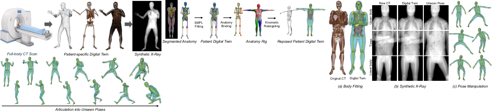
Figure 1: Overview. A single full-body CT scan is converted into a patient-specific articulated digital twin, permitting unseen pose manipulation and corresponding synthetic radiograph rendering.
Methodology
The pipeline consists of three key stages: patient anatomical modeling, anatomy-aware kinematic binding, and pose retargeting.
Patient Anatomical Modeling:
The anatomical model is constructed by segmenting the full-body CT using TotalSegmentator to extract bones and organ segmentation masks. Surface meshes are generated via Marching Cubes and Taubin smoothing. To impart articulation capability, a parametric SMPL human body model is fitted to the CT-derived geometry. Skeletal keypoint alignment initializes the fit, with a multi-term objective that prioritizes skeletal enclosure within the SMPL surface (computed via chamfer distance and segment inclusion losses), subject to anatomical priors and shape regularization. Unlike in previous work, the SMPL fit is driven to envelop the segmented patient skeleton, establishing a kinematic scaffold onto authentic patient geometry.
Figure 2: Overview of the pipeline, from CT segmentation to model fitting, kinematic binding, and pose retargeting.
Anatomy-Aware Kinematic Binding:
The segmented anatomical structures are assigned to a reduced kinematic tree rooted in the pelvis, with individualized joint anchors computed from CT mesh geometry. Rather than using mesh-based linear blend skinning as with standard SMPL animation, the method applies rigid transforms to each bone, preserving anatomical structural integrity during pose retargeting. Organs and non-bony soft-tissues are grouped and moved according to their attached skeletal segments, thus maintaining consistent internal organization during global pose changes.
Pose Retargeting:
New body configurations are specified using SMPL pose vectors, enabling arbitrary motion via the SMPL canonical pose space (e.g., sample sequences from the AMASS motion database). The rigid joint transformations computed from the difference in pose vectors are propagated to the CT-derived anatomy via forward kinematics, resulting in anatomically faithful repositioning of patient skeletal geometry, with organs following their associated segments.
Figure 3: (a) The fitted SMPL envelope encloses the CT skeleton; (b) DRRs from CT, digital twin at original pose, and articulated twin in new pose; (c) The articulated twin rendered in several unseen target poses.
Experimental Results
Quantitative evaluation was performed using three full-body CT datasets (NMDID). The models were assessed along three axes:
- Fitting Accuracy:
- Chamfer distance between fitted SMPL and CT surfaces achieved 15.8 ± 4.0 mm globally.
- Skeletal enclosure was 95.9 ± 1.8%, with the highest enclosure (99.0%) in the lower body.
- Localized fitting errors are attributed to the parametric SMPL model's inability to represent all soft-tissue anatomical variations, especially when inferring the standing pose from a supine-acquired body.
- DRR Recomposition Fidelity:
- Synthetic DRRs from recomposed twins (at the acquisition pose) preserve major radiographic structure.
- SSIM = 0.872 ± 0.016 and PSNR = 18.5 ± 1.4 dB across 72 paired projections.
- Structural fidelity is highest for skeletal regions; modest PSNR reflects residual soft-tissue fill limitations but does not affect critical bony alignment.
- Articulation Consistency:
- Across 28 sampled AMASS poses, anatomical enclosure after articulation remained at 94.4 ± 0.4%, i.e., no catastrophic breakdown or skeleton-envelope penetration.
- Qualitative DRRs of twins in novel poses demonstrate pose-dependent projection changes and retention of anatomical self-consistency.
Implications and Future Directions
This approach directly addresses the need for pose-controllable, patient-specific digital twins in CT-based simulation. It allows for arbitrary body pose manipulation while preserving radiographically meaningful anatomy, thus enabling new experiments for positioning studies, algorithm validation, and surgical workflow analysis with a single source CT scan per patient. In comparison to prior phantoms (e.g., XCAT-3.0 [dahal_xcat-30_2024]) or generative approaches (e.g., BOSS [shetty_boss_2023]), this method uniquely maintains full patient specificity throughout the musculoskeletal system while being pose controllable.
Certain limitations remain. The evaluation cohort is small (N=3) and downstream validation is currently restricted to structural enclosure and synthetic imaging. The model primarily implements rigid skeletal articulation; organ and soft-tissue motion is handled by co-segmental propagation without modeling non-rigid deformations, which may become a limitation in applications requiring internal organ mobility or deformation realism. Extreme SMPL poses occasionally incur local artifact, particularly in distal or non-canonical postures, reflecting limits of the SMPL fit to a static CT-derived shape. These limitations suggest future research in integrating advanced, patient-specific non-rigid modeling, automated keypoint annotation, and validation across larger, more heterogeneous populations.
Conclusion
This work demonstrates the feasibility of constructing articulated, patient-specific digital twins from a single CT acquisition, enabling pose-dependent anatomical and radiographic simulation. By leveraging kinematic priors from SMPL to impose articulation on CT-derived geometry, the method closes a significant gap in digital twin technology for medical imaging—enabling for the first time view- and pose-controllable patient models from existing clinical imaging data. The approach is positioned for extension into broader synthetic imaging, algorithm benchmarking, and advanced pre-interventional planning. Integration of non-rigid organ motion models and population-scale validation represents a natural next direction for continuing research on this topic.
Reference:
"Patient-Specific Articulated Digital Twins from a Single Full-Body CT Scan" (2607.02156)