- The paper presents a topology-optimized dielectric metasurface that achieves polarisation-switchable imaging between brightfield (identity) and phase-contrast (spatial derivative) modalities.
- It details a design process using inverse topology optimisation to engineer a nonlocal angular transfer function with robust diffraction-induced asymmetry and fabrication tolerance.
- Experimental validation on synthetic phase objects and biological samples confirms reliable quantitative phase extraction without additional optical components.
Overview
The paper "Multifunctional Imaging with an Inverse-Designed Nonlocal Metasurface" (2606.21051) introduces a topology-optimised metasurface enabling switchable brightfield and phase-contrast imaging at normal incidence via polarisation control, obviating the need for additional optical components or computational post-processing. This dielectric metasurface, fabricated using amorphous silicon on quartz, implements a nonlocal angular-filtering operator in the object plane, achieving two orthogonal imaging modalities: the identity operator for conventional brightfield (for x-polarised light), and a first-order spatial derivative for quantitative phase-contrast (for y-polarised light). The transfer function asymmetry central to phase contrast is engineered through diffraction effects and robustly achieved via pixel-wise topology optimisation. Experimental validation spans synthetic phase patterns and unstained biological samples, demonstrating the metasurface's computational and imaging efficacy.
Topology optimisation is employed to determine the binary permittivity distribution within the unit cell of the metasurface, targeting a polarisation-dependent angular optical transfer function (OTF). The OTF for x-polarisation is constant, yielding identity transmission (brightfield), while for y-polarisation, it is linear in kx, enabling spatial differentiation and thus phase contrast. Robustness to fabrication defects (dilation and erosion) is explicitly incorporated into the optimisation loop.
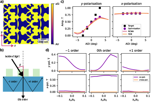
Figure 1: Final metasurface design and its simulated optical properties, showing period choice, asymmetric diffracted orders, simulated OTFs for both polarisations, and diffraction efficiency as a function of spatial frequency.
Diffraction-generated asymmetry is achieved by periodic structuring that allows coupling into non-propagating higher-order modes trapped by total internal reflection in the substrate. The device operates at 886 nm, with a design NA of 0.05, tuned for quantitative phase imaging of transparent objects. Contrast and slope of the OTF are controlled and validated via RCWA and FEM simulations, matching the required imaging modalities and demonstrating robustness for small geometrical variations.
Fabrication and Characterisation
The metasurface is fabricated via electron beam lithography followed by pattern transfer into amorphous silicon, producing an ultra-thin, freeform structure compatible with standard photodetector technologies. HIM micrographs confirm design fidelity, with only minor non-uniform dilation and negligible impact from the thin Al2O3 etch mask, as shown by simulated and measured transmission spectra.
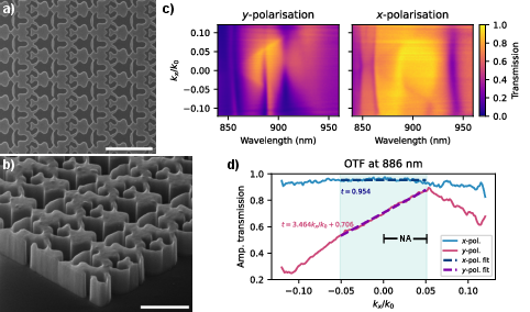
Figure 2: HIM images of the fabricated metasurface, transmission spectrum as a function of spatial frequency, and experimentally measured OTF for 886 nm illumination.
Experimental characterisation using back focal plane spectrometry yields OTFs matching the design: t=0.954±0.006 for x-polarisation (identity), and t=(3.464±0.150)kx/k0+(0.706±0.005) for y0-polarisation (linear phase gradient transfer). Operational wavelength is slightly blueshifted versus simulation due to minor fabrication errors.
Imaging Experiments: Synthetic Phase Objects
Using greyscale lithography, phase targets with controlled excursions are fabricated and imaged with the metasurface in a custom microscope setup. For phase objects, only rapidly varying features are visible in unprocessed images; the metasurface enables pseudo-3D visualization of phase gradients along y1, with high fidelity recovery compared to analytical ground truth (RMSE y2 rady3).
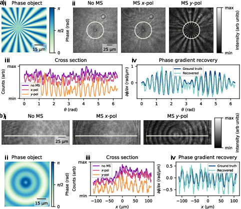
Figure 3: Imaging experiments on synthetic phase objects, including AFM-derived phase maps, intensity images with and without metasurface, cross-sections, and quantitative recovered phase gradient profiles.
Diffraction artifacts from metasurface edges and fabrication defects are minimized via averaging and robust design. The device quantifies the phase gradient directly from intensity images, using measured OTF coefficients and quadratic inversion, with tilt correction optimized for each experiment.
Biological Imaging Application
Imaging of unstained HeLa cells and human ovarian cancer tissue is performed with the metasurface placed in the object plane. Brightfield imaging yields low contrast; phase-gradient mode via y4-polarisation and subsequent gradient recovery reveals cell morphology and tissue microstructure, matching DIC reference images, albeit with lower spatial resolution (NA = 0.05 vs NA = 0.4 for DIC).
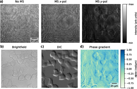
Figure 4: HeLa cell imaging with metasurface, showing brightfield, phase-gradient, and DIC modalities, and recovered phase gradient mapping.
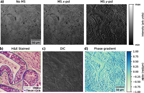
Figure 5: Imaging of unstained ovarian cancer tissue, with enhanced visualisation of intra-cystic spaces and cell morphology via metasurface, compared to brightfield, DIC, and stained reference images.
Averaging over multiple offsets reduces artefacts, leveraging the nonlocal uniformity of angular response. The metasurface enables label-free imaging and pseudo-3D phase-contrast directly in the object plane, with the operational modality swapped via simple polarisation rotation.
Algorithmic and Practical Implications
The topology optimisation workflow integrates symmetry enforcement, minimum feature size, binarisation, and robust variant (dilation/erosion) averaging, coupled to RCWA electromagnetic solvers and ADAM gradient descent.
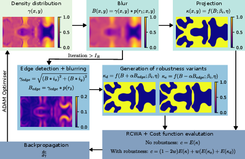
Figure 6: Flowchart of the topology optimisation algorithm, showing robustness loop for fabrication defect tolerance.
Robustness analysis confirms operational stability across realistic defect scales, with broad linear OTF regime and negligible impact on brightfield mode.
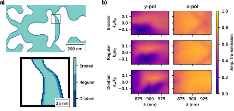
Figure 7: Overlaid robustness variants and OTF versus wavelength, demonstrating device reliability against fabrication imprecision.
Comparison between fabricated and designed geometries highlights non-uniform dilation as principal source of operational blueshift, suggesting future integration of stochastic defect models and further process calibration.
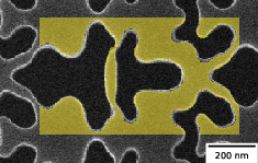
Figure 8: Overlay of designed and fabricated metasurface geometries, indicating regions of dilation and agreement.
Theoretical Impact and Future Directions
The polarisation-switchable metasurface advances the field of nonlocal, object-plane optical computation, enabling compact, real-time phase imaging without mechanical movement, additional optics, or multi-shot acquisition. The approach is extendable to broadband or multi-modal devices, with potential for tailored OTFs for target-specific imaging or object recognition. Topologically optimised metasurfaces offer precise control over NA and computational function, facilitating integration into both object and image plane systems, including computational cameras.
Further algorithmic enhancements could include stochastic modelling of fabrication, integration with scalable mass-production lithography, and development of multi-gradient recovery schemes enabling full quantitative phase imaging. Extension to broadband operation would enable low-cost, label-free imaging with LED sources.
Conclusion
This research establishes a compact, switchable imaging metasurface capable of real-time brightfield and phase-gradient microscopy via polarisation control. Topology optimisation allows robust transfer function engineering, implemented with diffraction-driven asymmetry at normal incidence. Experimental results demonstrate the device's ability to facilitate quantitative optical computation and enhance visualisation of transparent biological samples without the need for auxiliary components. This paradigm paves the way for ultra-compact, low-power, label-free imaging platforms with tunable computational functionality.