- The paper introduces a projection-based methodology to marginalize nuisances such as optode coupling, RONI absorption, and baseline misspecification.
- It leverages Monte Carlo simulations and singular vector analysis to construct efficient projections that reduce artifacts in DOT reconstructions.
- Bayesian inversion with these projections achieves robust ROI reconstructions, significantly lowering L2 errors under nonideal conditions.
Projections for Handling Uncertainties and Enabling Domain Truncation in Diffuse Optical Tomography
Introduction
The paper "Projections for handling uncertainties and enabling domain truncation in diffuse optical tomography" (2604.26548) presents a projection-based methodology for mitigating the effects of modeling inaccuracies in diffuse optical tomography (DOT). The authors specifically address three sources of model uncertainty: changes in optode coupling coefficients, absorption variations outside the region of interest (ROI), and misspecification of tissue baseline optical parameters. The rationale is adapting the inversion algorithm to project the measurement model onto the orthogonal complement of subspaces associated with nuisance parameters, thereby marginalizing their influence on reconstructed absorption changes. This essay delineates the methodological constructs, numerical findings, and the broader implications in functional neuroimaging.
DOT Forward Model and Linearization
DOT estimates spatially resolved absorption coefficient changes correlated with hemodynamic activity by measuring the amplitude and phase shifts of modulated light after propagation through tissue. The authors employ Monte Carlo (MC) simulations to model photon transport in a voxelized neonatal head, accurately capturing complex domain geometries and tissue heterogeneities.
The primary linearized forward model considered is
y=Jxtotal+e=Jx+J~x~+e,
where y are measurement differences, x is the ROI absorption change, and x~ represents changes in the region of non-interest (RONI). Jacobians J and J~ relate these changes to measurements, respectively. A similar Jacobian is defined for coupling coefficients, denoted Jc. Linearization is justified for small-to-moderate perturbations, given the underlying nonlinearity of the forward problem.
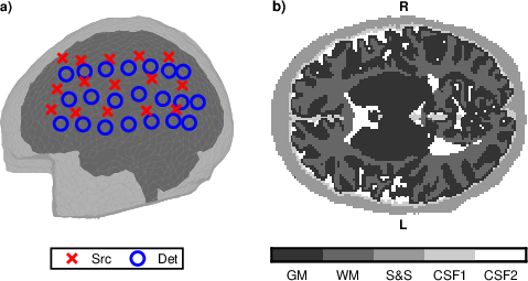
Figure 1: The optode configuration and segmentation of the neonatal DOT head model, with sources and detectors localized over the left hemisphere and tissue types highlighted.
Projection Construction for Nuisance Marginalization
The central innovation is the systematic marginalization of nuisance parameter influence via orthogonal projections:
- Low-Dimensional Nuisance Parameters (Coupling Coefficients): The range of Jc is marginally low-dimensional; the projection
P=I−Jc(Jc⊤Jc)−1Jc⊤
is applied to both data and the forward operator, cleanly separating the effects of coupling coefficient perturbations.
- High-Dimensional Nuisance Parameters (RONI Absorptions): For RONI, direct projection onto the full range of J~ would severely degrade the signal. Instead, the approach utilizes a truncated subspace determined by dominant left singular vectors of a weighted y0, guided by prior covariance structure.
- Misspecified Baseline Parameters: Here, the nullspace of the projection is the space spanned by principal left singular vectors of the (weighted) difference of Jacobians at two baseline parameterizations, handling uncertainty in tissue-class optical properties.
- Compound Projections: For scenarios involving multiple concurrent uncertainties, the basis vectors corresponding to all relevant nuisance subspaces are concatenated, allowing joint marginalization.
The mathematical framework is fully general and simulated using meticulous MCX-based Jacobian construction over the segmented anatomy.
Bayesian Inversion Under Projections
A hierarchical Bayesian linear Gaussian model is utilized for inversion, with priors on voxel absorptions and additive Gaussian noise. After projection, the posterior mean becomes
y1
which encapsulates the data projected to the nuisance-marginalizing subspace, thus reflecting uncertainty-aware reconstruction. The approach is statistically coherent and computationally efficient due to the preserved low dimensionality after projection.
Experimental Protocol
A neonatal head volume is simulated, with finely detailed segmentation into scalp/skull, two CSF compartments, GM, and WM. Optical parameters are taken from literature for near-infrared light. The optode geometry covers a cortex-proximal field, with 15 sources and 21 detectors yielding 210 measurement pairs. GPU-accelerated MC guarantees high-fidelity measurements and efficient replay-based Jacobian computation.
Numerical Results
Reference Reconstructions
Initial reconstructions under idealized (mismodel-free) conditions yield low y2 errors (0.185/0.266 for the two targets), serving as a benchmark.
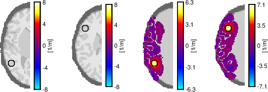
Figure 2: Representative cross-sections of target 1 and its reference L2 error reconstruction.
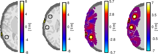
Figure 3: Representative cross-sections of target 2 and its reference L2 error reconstruction.
Mismodeled Coupling Coefficients
Simulations with randomized coupling coefficients (amplitude and phase) demonstrate that naive reconstructions become dominated by artifacts (y3 errors ≫ 1). Projections based on y4 restore the reconstructions to near-baseline quality (y5 errors 0.195/0.266), substantiating the approach's efficacy for low-dimensional nuisance marginalization.
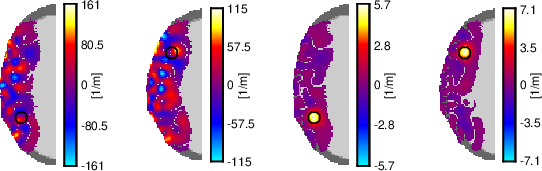
Figure 4: Comparison of reconstructions for target 1 without vs. with coupling projection; substantial artifact removal is observed.
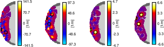
Figure 5: Projection-based reconstruction for target 2, showing artifact suppression.
RONI Absorption Marginalization
Domain truncation, where ROI excludes substantial tissue volumes, introduces interface artifacts if not handled. Projections onto subspaces determined by dominant eigenvectors of y6 effectively suppress these, with only marginal y7 error increases in the ROI (e.g., from 0.136 to 0.183).
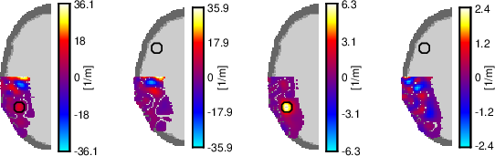
Figure 6: Domain truncation (top-half removed) and artifact-elimination in target 1 using RONI projection.
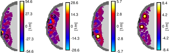
Figure 7: Artifact removal in brain-only reconstructions for target 2 when projecting out scalp/skull contributions.
Misspecified Baseline Absorption
Reconstructions with significantly erroneous baseline y8 for GM/WM change dramatically, introducing bias and loss of sensitivity. By projecting onto the singular vector subspaces of the difference Jacobian (between baseline and an alternative plausible value), a substantial fraction of the error is recovered (y9 reduced from over 1.1 to ≈0.39).
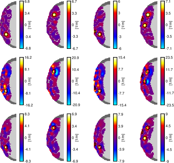
Figure 8: Multilevel reconstruction quality under correct, misspecified, and projected baseline absorption in both targets.
Simultaneous Uncertainties
Compound cases (nonideal coupling and RONI perturbations combined) affirm that joint projection removes even more severe artifacts, resulting in reconstructions whose x0 error is largely unaffected compared to single-nuisance scenarios.
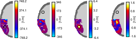
Figure 9: Joint nuisance (coupling + RONI) marginalization in target 1, yielding clean ROI reconstructions.
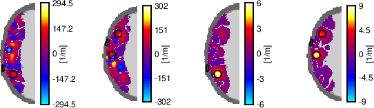
Figure 10: Compounded projection for target 2: clear suppression of multiple classes of modeling errors.
Implications and Future Directions
This projection-based strategy provides a rigorous, computationally tractable solution to primary sources of model mismatch in DOT and potentially other modalities with linearizable forward models and separable nuisance parameters. Critically, the approach bypasses the sampling-intensive methods such as the approximation error (AE) framework and avoids the computational bottleneck of deep learning or extensive MC marginalization.
Theoretical implications: The method generalizes Jacobian-based marginalization to arbitrary nuisance parameter structures, is compatible with high-dimensional Bayesian inversion, and provides a statistical mechanism to formalize truncated domains without explicit modeling of all tissue regions. The projection methodology is extensible to other inverse problems, e.g., EIT, and potentially multi-modal imaging.
Practical implications: Significant computational savings and robustness are enabled for longitudinal and real-time DOT reconstructions, as ROIs can be adaptively chosen with minimal loss of information. This is crucial for clinical settings involving neonates or moving subjects and where fast feedback is needed.
Future directions: Extension to scattering and anisotropy uncertainty, systematic optimization of the number and type of projected singular vectors, and data-driven selection of projection subspaces could further improve robustness. The impact of nonlinearities in high-contrast regimes, and integration with adaptive experimental design, remain open avenues.
Conclusion
The paper establishes that targeted orthogonal projections of the linearized forward model can marginalize specific modeling uncertainties in DOT reconstructions, achieving demonstrable reduction in artifacts and insensitivity to incomplete or uncertain modeling assumptions. The methodology is readily applicable in high-dimensional Bayesian settings, and the presented numerical results indicate clear superiority over naive inversion in the presence of nuisance parameters. This projection-based paradigm may catalyze improved accuracy, computational tractability, and adaptivity in next-generation functional optical imaging and beyond.