- The paper demonstrates that YOLOv12, paired with Otsu thresholding, achieves up to 99.3% test accuracy in multiclass AML cell classification.
- It uses dual segmentation strategies (hue and Otsu) to isolate both cell and nucleus features, enhancing signal-to-noise ratio for detection.
- Experimental results show YOLOv12 outperforms classic models like ResNet50, enabling rapid and reliable diagnostic suggestions in hematopathology.
Early Detection of Acute Myeloid Leukemia (AML) with YOLOv12: Methods and Empirical Evaluation
Introduction
This paper investigates the application of the YOLOv12 object detection architecture for the automated, multiclass classification of acute myeloid leukemia (AML)-related blood cell types. AML is a hematological malignancy with clinical urgency for rapid, accurate diagnosis due to its aggressive progression. Morphological differentiation of neoplastic myeloblasts from normal or other abnormal blood and bone marrow cells remains a practical challenge with high intra- and inter-observer variability. The research aims to address prevalent limitations in existing DL-based AML classification methods, namely poorly balanced datasets, suboptimal data curation, and reliance on outdated feature extraction techniques. The proposed workflow leverages two complementary segmentation strategies (Hue channel and Otsu thresholding), targeting both cell and nucleus structures, in combination with advanced transfer learning using YOLOv12, ResNet50, and Inception-ResNet50 v2.
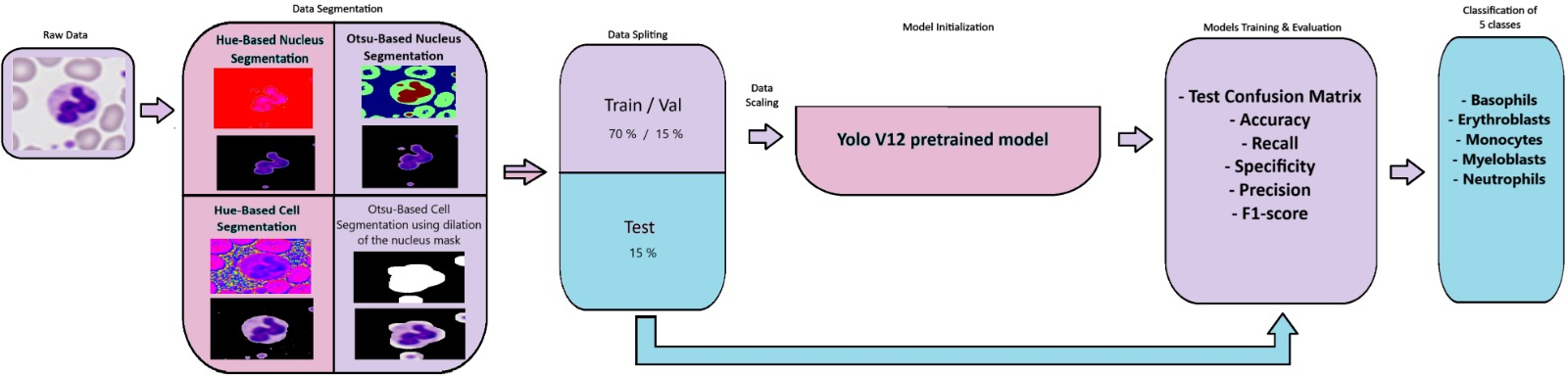
Figure 1: The implementation process of the proposed framework, spanning data preprocessing, segmentation, model training, and classification.
Datasets and Preprocessing
The dataset, comprising 5,000 annotated microscopic images, was curated from public sources including TCIA and Mendeley Data. It contains five diagnostically relevant classes: myeloblasts, segmented neutrophils, basophils, monocytes, and erythroblasts. Images were standardized (1024x1024, 24-bit RGB, Wright-Giemsa staining, magnified at 1000x) and divided into training, validation, and test partitions (70%, 15%, 15% respectively). To optimize discriminative feature extraction, both cell-level and nucleus-level foregrounds were isolated using either hue-based segmentation or Otsu's adaptive thresholding. Each of these four segmentation modalities supported subsequent DL-based classification model training.
YOLOv12 Architecture and Area Attention
YOLOv12, the latest evolution in the YOLO family, couples single-stage object detection with a novel area attention module designed to enhance feature context extraction without incurring prohibitive computational cost. Specifically, YOLOv12 implements area attention by partitioning feature maps into long horizontal or vertical strips (with default l=4), drastically reducing the attention computation complexity from O(2n2hd) to approximately O(21n2hd) while maintaining spatial acuity.
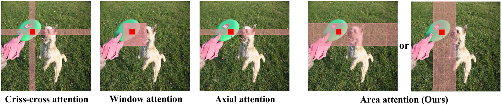
Figure 2: Area Attention divides the feature map into l equal vertical or horizontal segments for efficient and context-rich attention.
YOLOv12-N, the network variant selected for these clinical experiments, achieves a mean Average Precision (mAP) of 40.6% with a latency of 1.64 ms per image on T4 hardware, outperforming preceding YOLO iterations in both speed and accuracy.
Separate YOLOv12 models were trained and evaluated for each segmentation scenario (cell-hue, cell-otsu, nucleus-hue, nucleus-otsu). Performance was assessed using standard classification metrics: accuracy, sensitivity, specificity, precision, and F1-score, all derived from confusion matrices constructed at test time for each experimental branch. The study strictly avoids validation/test set cross-contamination during hyperparameter tuning.
Results
On hue-segmented cell images, YOLOv12 exhibited stable convergence with test/validation accuracy at 98.8%.
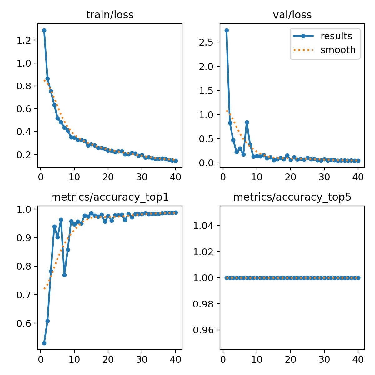
Figure 3: Training and validation loss for hue-segmented cell images processed by YOLOv12, showing steady decline and stabilization.
Classification confusion for cell types, as detailed in the confusion matrix, demonstrates high inter-class discriminability and minimal misclassification.
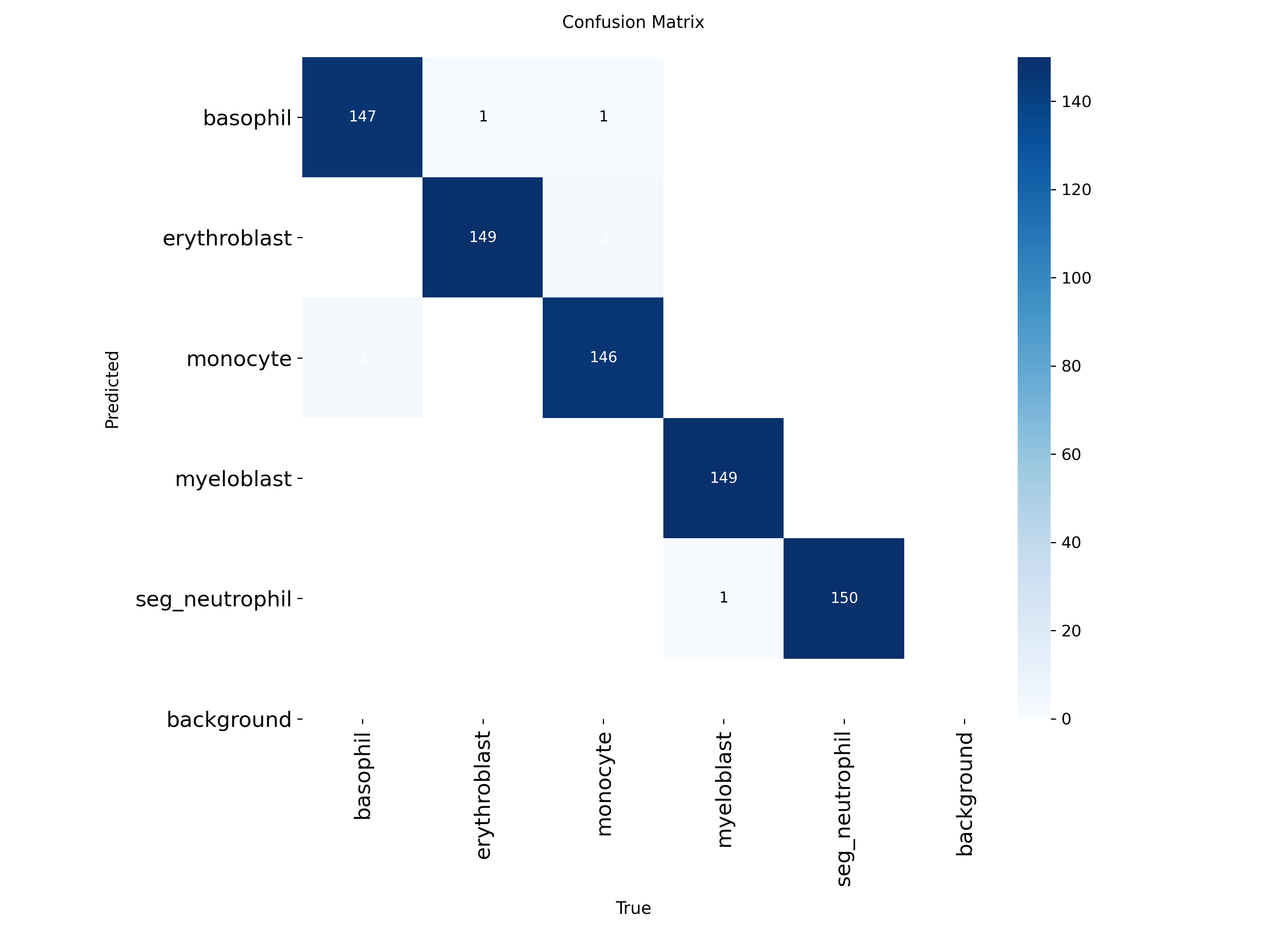
Figure 4: Confusion matrix for hue-segmented cell image classification, illustrating robust separation across the five phenotypes.
Cell image segmentation with Otsu thresholding yielded the optimal results, achieving both validation and test accuracy of 99.3%. Training and validation losses reflected rapid convergence with limited overfitting.
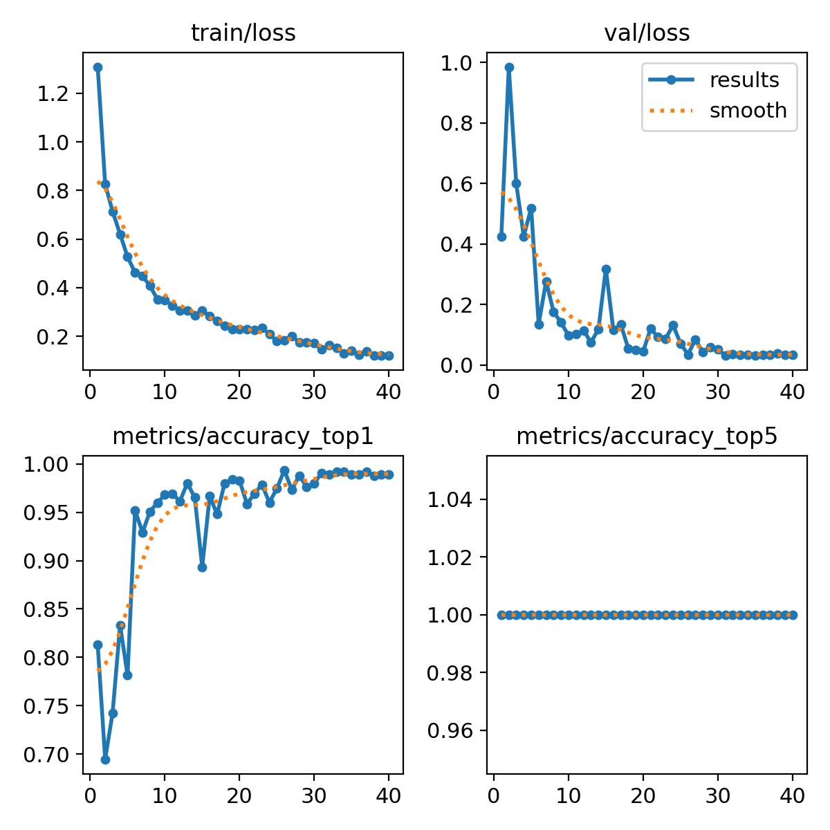
Figure 5: Training and validation loss for Otsu-segmented cell images processed by YOLOv12, supporting superior generalization.
The corresponding confusion matrix shows negligible off-diagonal errors, confirming the segmentation-model synergy.
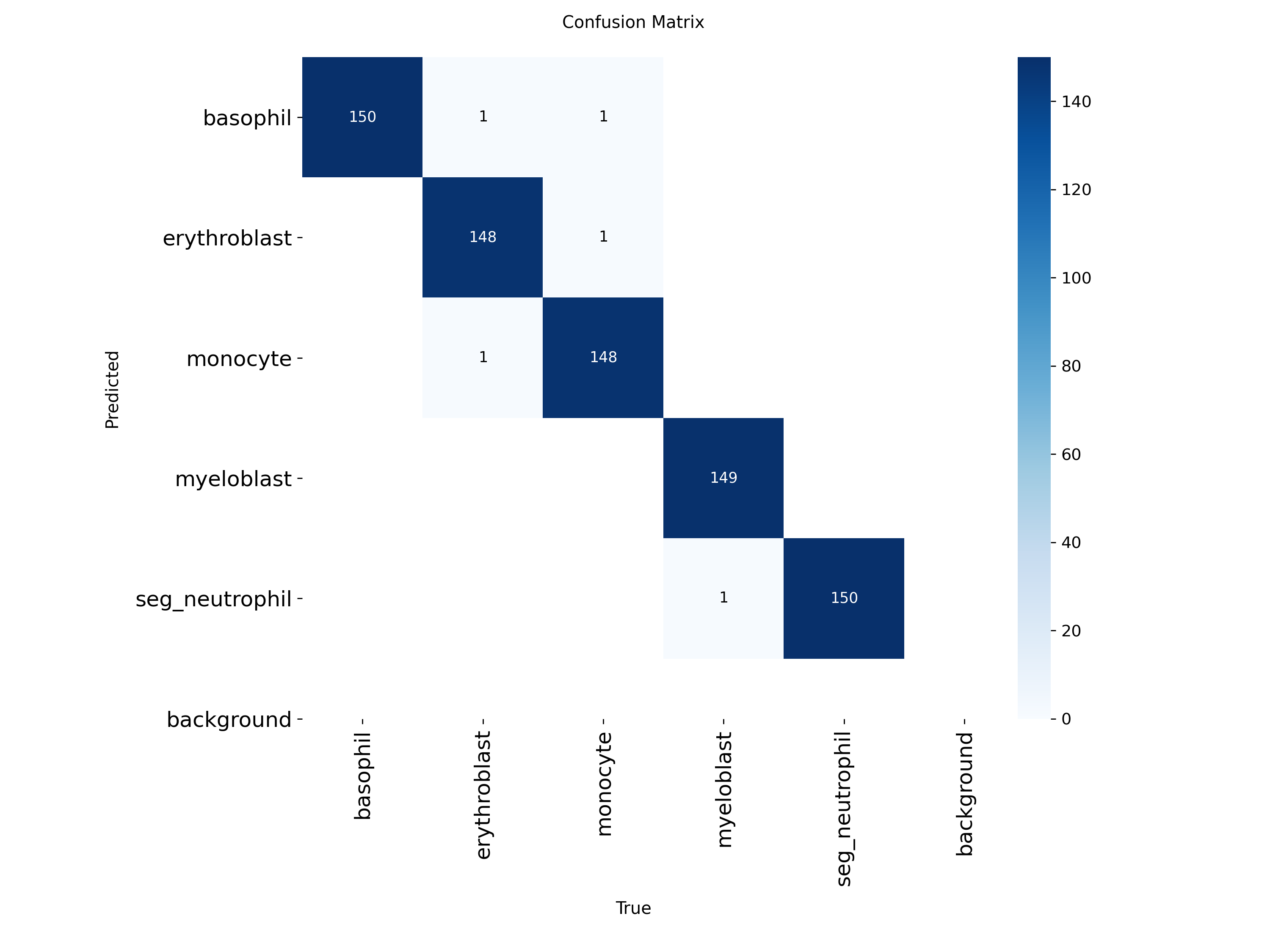
Figure 6: Confusion matrix for Otsu-thresholded cell image classification, indicating near-perfect prediction reliability.
Similar analyses on nucleus-based segmentations resulted in consistent validation/test accuracy of 98.8%, regardless of segmentation approach.
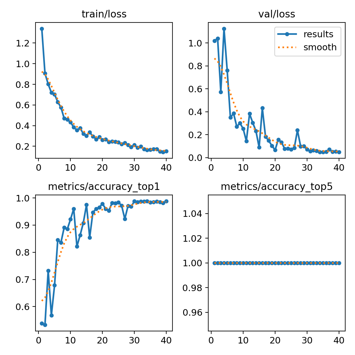
Figure 7: Loss curves for hue-segmented nucleus images with YOLOv12, with smooth convergence and minimal fluctuation.
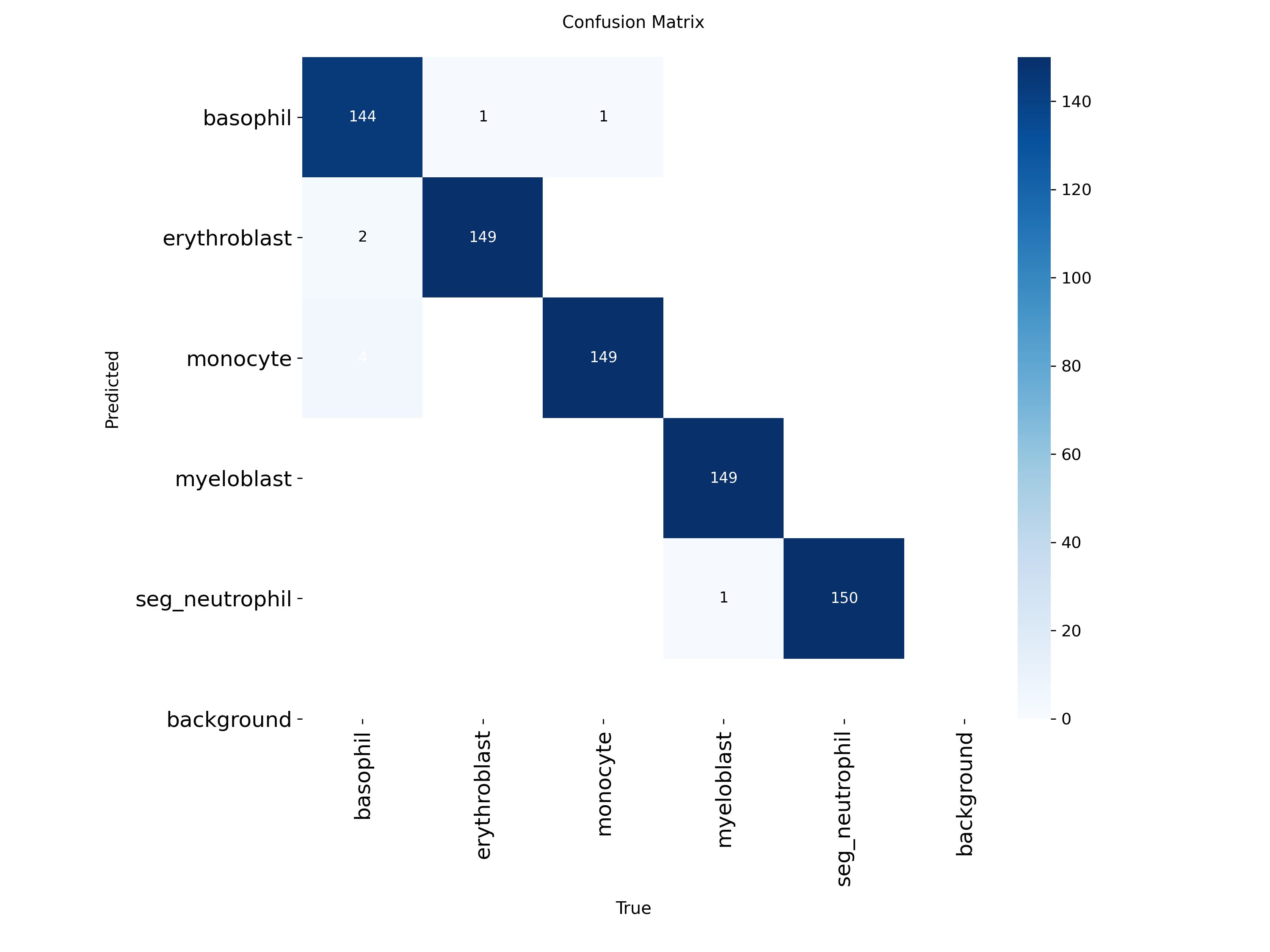
Figure 8: Confusion matrix for hue-segmented nucleus images, affirming multi-class reliability for subcellular structures.
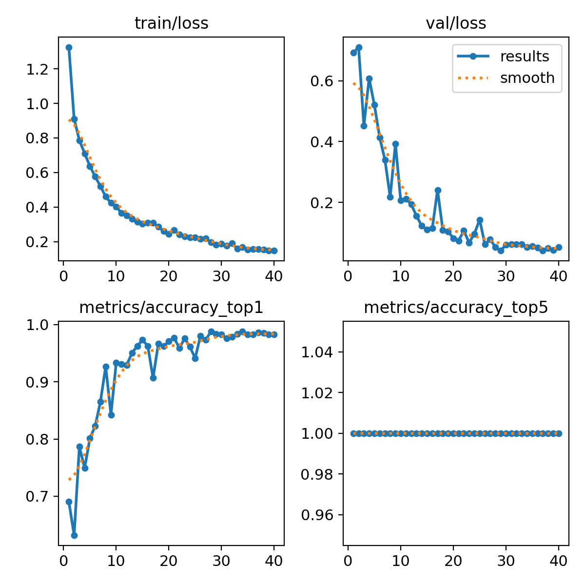
Figure 9: Loss curves for Otsu-segmented nucleus images by YOLOv12, showing stable reduction in loss.
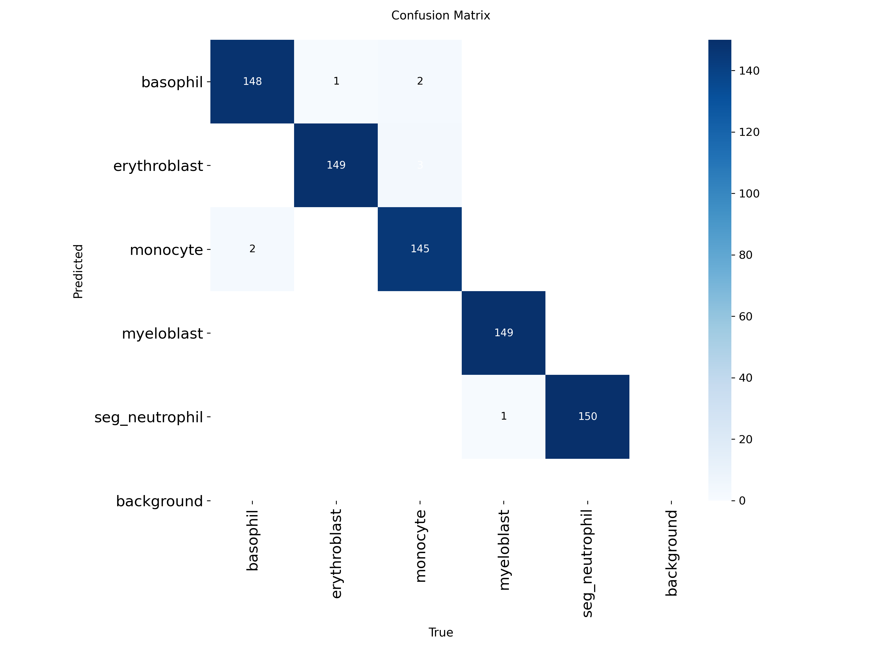
Figure 10: Confusion matrix for Otsu-thresholded nucleus image classification, confirming high specificity and sensitivity.
Comparative Model Analysis
Across all tasks, YOLOv12 consistently outperformed both ResNet50 and Inception-ResNet50 v2 baselines, establishing superiority in multiclass discrimination of AML-relevant cell phenotypes. The strongest numerical result reported is a 99.3% test accuracy on cell images segmented with Otsu thresholding—a statistically significant finding considering inter-dataset variability and annotation ambiguity in hematopathological samples.
Discussion
The experimental results underscore several core claims:
- Balanced and meticulously preprocessed datasets yield clinically relevant, generalizable models for AML subtype recognition.
- Cell-level segmentation with optimal adaptive thresholding substantially enhances the signal-to-noise ratio for morphological classifier input, outperforming both nucleus-focused and color-only (hue) segmentation pipelines.
- The area attention module in YOLOv12 provides scalable context aggregation required for robust feature representation in high-dimensional histopathological domains, without the inference-time penalties observed in alternative transformer or vision-attention architectures.
- The observed accuracy improvements relative to prior AML-focused DL methods (cf. (2604.16082)) highlight the benefit of a harmonized preprocessing, segmentation, and detection pipeline utilizing modern single-stage detectors.
Implications and Future Perspectives
The integration of advanced real-time object detectors with targeted pre-segmentation poses significant opportunity for both practical and translational hematopathology. The described framework supports rapid, expert-level, reproducible classification of AML-related cell morphotypes, and could be deployed as an assistive diagnostic tool for early intervention workflows, especially in resource-limited clinical contexts. The high-confidence classification potential can further support downstream tasks such as subclone tracking, blast quantification, or rare phenotype discovery.
Future research avenues may include:
- Expanding the cell class ontology to cover rarer AML variants and cytogenetic subtypes.
- Integrating multi-modal data (cytomorphological, immunophenotypic, molecular) to further disambiguate challenging cases.
- Investigating self-supervised pretraining or prompt-based learning, exploiting larger unlabeled microscopy corpora.
- Validating the pipeline in prospective, multi-center clinical trials with real-world whole-slide imaging data.
Conclusion
The study rigorously demonstrates that YOLOv12, coupled with Otsu-thresholded cell segmentation, delivers state-of-the-art multiclass classification of AML-essential blood cell types. The findings provide principled guidance for future automated hematopathology solutions, confirming the merit of modern attention-based single-stage detectors when combined with carefully selected preprocessing pipelines. This architecture sets a benchmark for future efforts in explainable, deployable, and scalable leukemia diagnostic systems.