- The paper demonstrates a ResNet-based CNN with patch-wise classification achieving over 0.80 Dice coefficient for accurate retinal cyst segmentation.
- It employs robust data augmentation and vendor-agnostic preprocessing to consistently outperform traditional segmentation methods.
- The approach improves clinical reproducibility in CME assessment and offers a flexible framework for other medical imaging challenges.
Retinal Cyst Detection from Optical Coherence Tomography Images: A ResNet-Based Approach
Introduction
Retinal cystoid macular edema (CME) represents a critical vision-threatening manifestation in multiple ocular pathologies, including diabetic retinopathy and age-related macular degeneration. The identification and quantification of intraretinal cysts from optical coherence tomography (OCT) images is essential for guiding therapeutic decisions and monitoring disease progression. However, achieving robust and accurate segmentation remains challenging across heterogeneous imaging platforms, primarily due to pronounced speckle noise and variability in scanner quality. The present work introduces a ResNet-based convolutional neural network (CNN) utilizing patch-wise classification for automated segmentation and volumetric quantification of retinal cysts, evaluated using the OPTIMA challenge dataset spanning four commercial OCT devices.
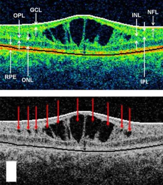
Figure 1: Upper panel depicts segmented intra-retinal layers with visible cysts in OCT; lower panel marks true cystic regions in the ground truth image.
Prior methodologies for cyst segmentation encompass threshold-based image processing, statistical learning, deformable models, denoising techniques, classical machine learning classifiers (e.g., random forest, AdaBoost), and graph-based approaches. Most of these methods demonstrate acceptable performance on high-quality (e.g., Spectralis) OCT scans but degrade considerably when confronted with lower-quality vendor images, such as those from Topcon.
Feedforward and feedback (recurrent) artificial neural networks have been applied in prior work, but their inability to mitigate vanishing gradient issues in deeper architectures limits segmentation fidelity. Residual neural networks (ResNets) introduce skip connections that preserve information flow, enabling the training of substantially deeper networks with enhanced segmentation accuracy and data generalization.
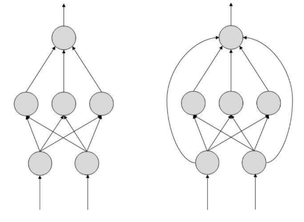
Figure 2: Feedforward network topology illustrating unidirectional information flow through consecutive layers.
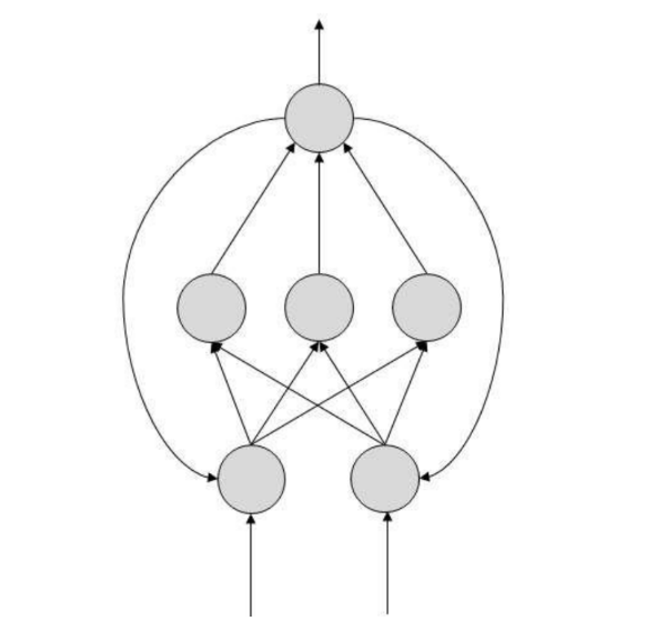
Figure 3: Feedback network with recurrent cycles, supporting temporal and sequence modeling.
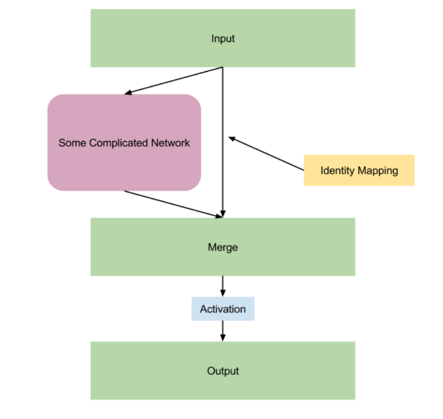
Figure 4: Information flow in a residual network, demonstrating improved propagation through identity mapping.
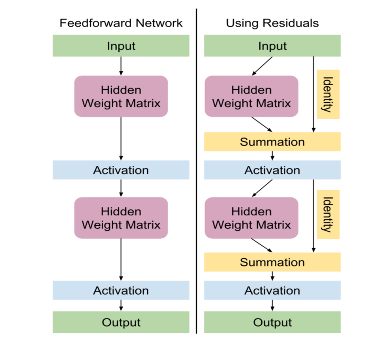
Figure 5: Comparison of feedforward and residual network architectures underlying information preservation.
Methodology
Data Preprocessing
The multi-vendor OPTIMA challenge dataset, comprising 3D OCT scans, was decomposed into 2D images. Only intra-retinal regions, as delimited by the Iowa Reference Algorithm, were retained. Images were denoised using non-local means filtering and their contrast enhanced via CLAHE. After resizing to 256×512 pixels, images were divided into non-overlapping 11×11 patches. The ground truth for each patch was assigned by its central pixel classification using consensus segmentation from two graders.
Model Architecture and Training
A vanilla ResNet18 architecture was configured without transfer learning. The training regime followed strong data augmentation (random rotations, flips, scale shifts), categorical cross-entropy loss, and the Adam optimizer (learning rate = 3×10⁻⁴, β1=0.9, β2=0.999). Training proceeded for 100 epochs with a batch size of four.
For pixel-wise segmentation during inference, overlapping patches were classified and reassembled to reconstruct the cyst mask for each image.
Evaluation and Results
Performance was measured using the Dice coefficient, sensitivity (recall), and precision, with reference to multi-grader ground truth masks. The ResNet-based approach achieved mean Dice coefficients exceeding 0.80 across all vendors, outperforming previous state-of-the-art algorithms by a substantial margin (68% Dice in prior art).
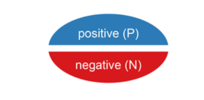
Figure 6: Two ground-truth classes (cyst, non-cyst) present as observed labels within the test dataset.
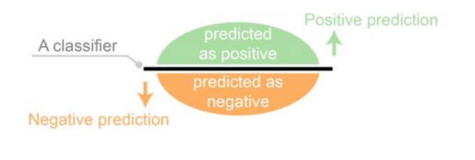
Figure 7: Predicted classes of an ideal classifier, showing perfect agreement with the ground truth.
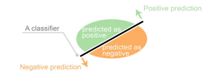
Figure 8: Predicted classes of the trained classifier with partial agreement to actual labels due to errors.
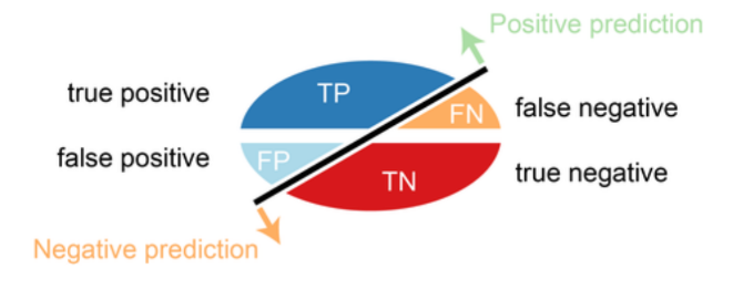
Figure 9: Four canonical outcomes for a classifier: true positive, true negative, false positive, and false negative.
The model demonstrated strong cross-vendor generalization, evidenced by:
- Average Dice coefficient: 0.82±0.08
- High precision (>0.98) and robust recall
- Consistent performance on low-quality (Topcon) images, unlike earlier methods
Comparative analysis against legacy methods (e.g., thresholding, deformable models, region flooding with texture analysis, random forest classifiers) reveals superior segmentation accuracy and reduced sensitivity to vendor-specific noise.
Discussion and Implications
The patch-wise ResNet strategy exhibits several key properties:
- High segmentation accuracy sustained across vendors and diverse image noise profiles
- Effective volumetric quantification of cysts suitable for clinical use in CME assessment
- Minimal reliance on manual parameter tuning or vendor-specific preprocessing
Implications are twofold:
Practically, the method enhances the reliability and reproducibility of CME severity estimation, which can be directly integrated into ophthalmological clinical workflows for diagnosis, monitoring, and outcome prediction.
Theoretically, the demonstrated vendor-agnostic generalization affirms the effectiveness of deep residual connections in mitigating representation loss and vanishing gradients within biomedical image segmentation. The methodology provides a template for future architectures targeting other high-variability medical imaging tasks.
Future Directions
Several vectors for advancement are evident:
- Investigation of advanced denoising pipelines, alternative patch/block sizes, and transfer learning with large-scale image datasets (e.g., ImageNet)
- Benchmarking against novel architectures including UNet and GoogleNet for further improvement in segmentation robustness
- Expansion to 3D and temporal domain (3D+T) for tracking cyst evolution, harnessing generative or autoregressive architectures
- Integration of domain-specific frontends or graphical user interfaces for streamlined clinical deployment
- Embedding the segmentation module within multi-agent clinical decision support systems, analogous to agentic IR and recommender systems
- Implementation of privacy-preserving mechanisms such as domain-specific steganographic encoding for protected health data handling
Conclusion
The proposed ResNet-based patchwise classification framework establishes a new benchmark for intra-retinal cyst segmentation in OCT imagery. It delivers robust, cross-vendor, high-fidelity segmentation, enabling accurate volumetric quantification with minimal human intervention. The technical paradigm validated in this study offers a flexible foundation for generalization to other clinical image analysis applications and informs the roadmap for deploying AI-enabled diagnostic tools in ophthalmic practice.
Reference:
"Retinal Cyst Detection from Optical Coherence Tomography Images" (2604.10843)