- The paper introduces VCC-DSA, a novel model that suppresses motion artifacts in DSA imaging using a vascular consistency constraint.
- It utilizes a multi-scale residual dense network with LSMP to extract fine vessel details and reduce artifact interference.
- Extensive evaluations demonstrate that VCC-DSA outperforms baselines in PSNR and SSIM while generalizing across heterogeneous datasets.
VCC-DSA: A Vascular Consistency-Constrained Imaging Model for Robust DSA Artifact Suppression
Digital Subtraction Angiography (DSA) remains the clinical reference standard for cerebrovascular imaging but is hampered by motion artifacts arising from patient movement and physiological processes. Traditional registration-based compensation is fundamentally limited by the inability to accurately model three-dimensional patient motion in a two-dimensional projection framework, resulting in residual artifacts that impede precise vascular visualization and diagnostic utility. Learning-based methods that treat DSA synthesis as a style transfer task are similarly constrained by training data ill-posedness: anatomical structures with similar density profiles (e.g., bone and vasculature) confound the extraction of authentic vascular signals, especially in the absence of robust, artifact-free ground truth labels and paired live-masked images (Figure 1).
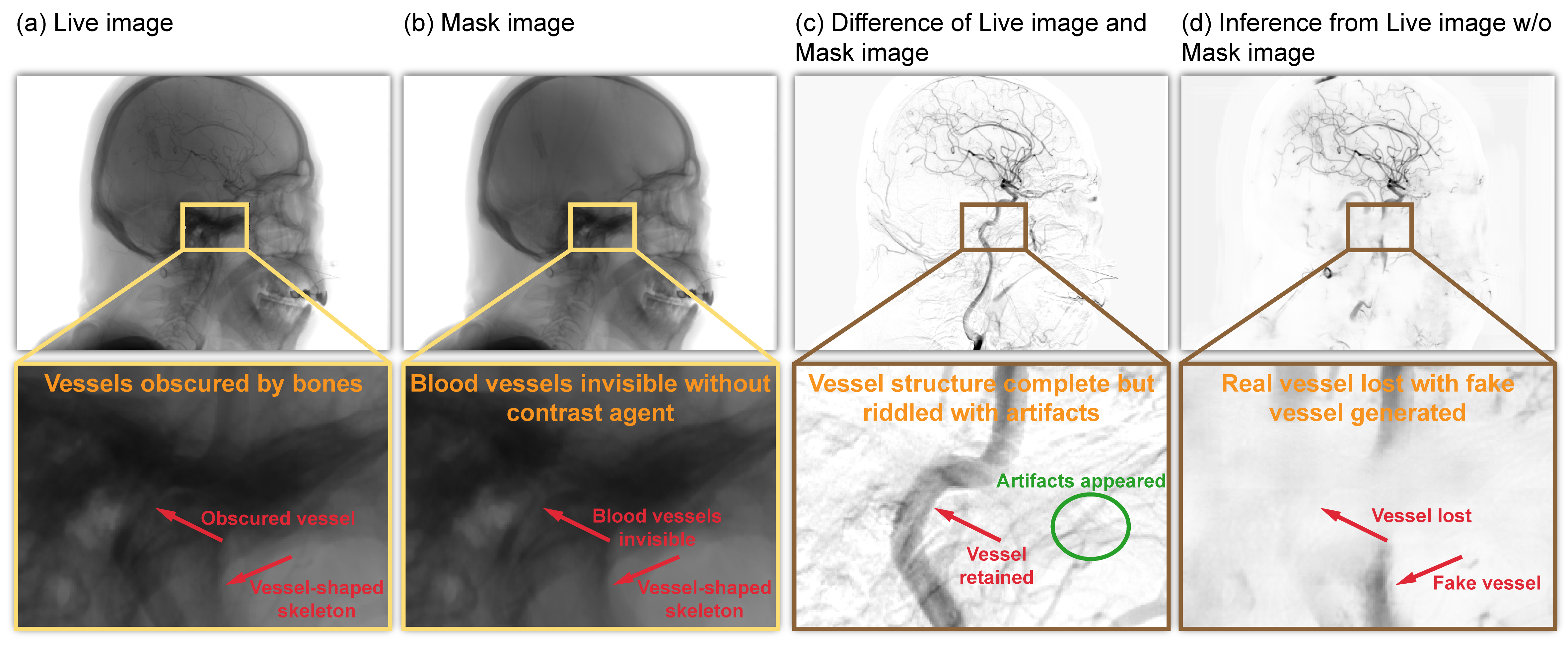
Figure 1: DSA synthesis is challenged by low vessel/background contrast, artifact-rich subtractions due to motion, and ambiguity in structure attribution in data.
Model Architecture and Design Rationale
The VCC-DSA framework introduces a holistic approach tailored to the simultaneous enhancement of vascular signal and robust suppression of motion artifacts. It is driven by four synergistic components:
- Learning-based Subtraction Mapping Paradigm (LSMP): Instead of inferring vessels directly from the live image, LSMP couples mask (pre-contrast) and live (post-contrast) images as input to model vascular structure as the information missing in their difference, explicitly leveraging background contextual cues and addressing the mapping ill-posedness.
- Residual Dense Block (RDB) Network with Detail Shortcut: The backbone comprises a multi-scale encoder-decoder with RDBs for high-frequency detail retention and deep feature fusion, enabling the model to resolve fine vessel termini and small-caliber branches in complex anatomical regions (Figure 2, Figure 3).
- Vascular Consistency Strategy (VCS): By formulating a structural consistency constraint across multiple mask-live pairs with shared live input, the model implicitly learns vascular features invariant to motion-induced transformations, regularizing representation space to favor true vascular signals and de-emphasize movement-induced artifacts.
- Mixup-based Data Self-evolution Strategy (MDSS): Through dynamic augmentation of training data and label targets—combining DSA-like features from a learned "vascular bank" pool—MDSS further reinforces the extraction of consistent, artifact-suppressed vascular representations (Figure 4).
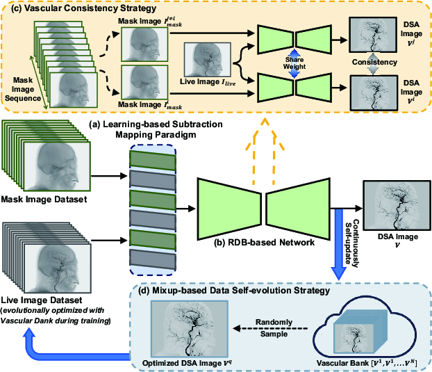
Figure 2: VCC-DSA overall architecture highlights LSMP, the RDB backbone, VCS, and MDSS components.
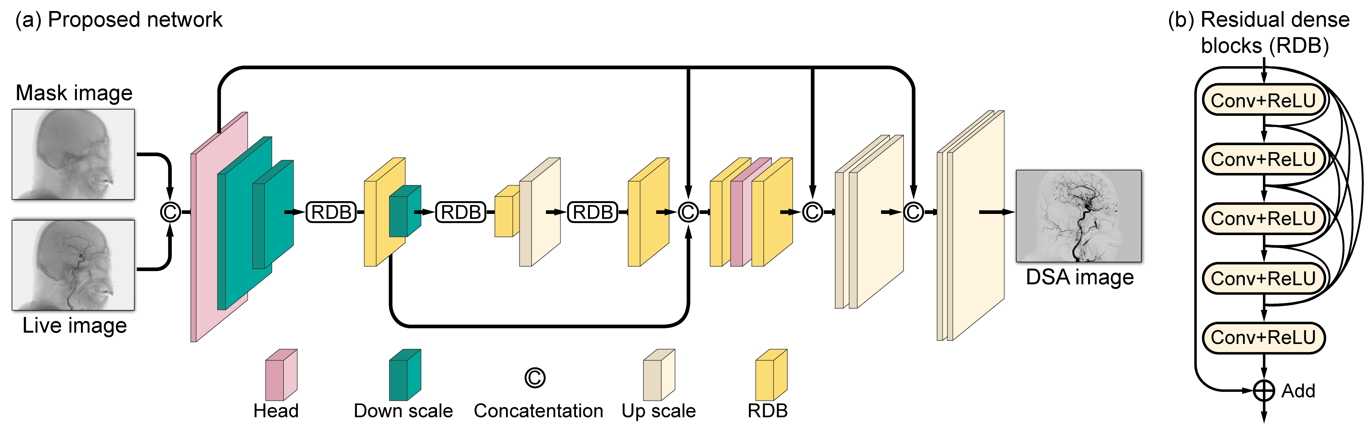
Figure 3: Detailed RDB network diagram, delineating multi-scale feature propagation and dense connectivity for robust detail preservation.
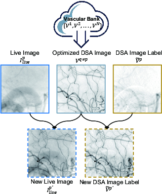
Figure 4: MDSS process: augmentation and optimization of training samples label-wise, evolving vascular clarity and boosting discriminative learning.
Training Objectives and Supervision
The combined objective function incorporates a fidelity term (guiding output towards label-similar appearance for vascular content) and a consistency term (minimizing difference between outputs from different mask-live pairings), weighted by a tunable scalar λ. Loss optimization ensures convergence to a solution that prioritizes vascular feature similarity across physiologic motion and label contamination. The MDSS procedure further ensures that label set evolution maintains high vascular fidelity while progressively reducing artifact content.
Experimental Evaluation
Datasets and Experimental Protocol
Experiments utilize both clinical human DSA datasets (United Imaging uAngio 960, Philips Azurion 7 B20) and a prospectively acquired, motion-free animal dataset (porcine model under general anesthesia with respiratory and mechanical immobilization). The latter allows precise quantitative benchmarking, circumventing the issue of artifact-contaminated ground truths prevalent in routine clinical data.
Quantitative and Qualitative Results
VCC-DSA demonstrates high-fidelity vascular reconstruction and robust artifact suppression, outperforming baseline methods by substantial margins in both PSNR and SSIM on the animal dataset (PSNR = 43.19 dB, SSIM = 98.8%). Compared to state-of-the-art learning-based and classical approaches, the VCC-DSA model produces DSA images with clearer vessel-to-background separation, better preservation of peripheral and terminal branches, and lower rates of false-positive vessel detection or artifact hallucination (Figure 5).
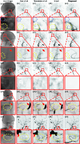
Figure 5: Qualitative DSA results; VCC-DSA (d) yields superior vessel clarity, eliminates oxygen tube and dental artifacts and suppresses blur.
Model generalizability is confirmed by robust cross-device and cross-species transfer. The method remains effective—and even superior in artifact suppression—when evaluated on unseen datasets and animal-to-human generalization scenarios (Figure 6, Figure 7).
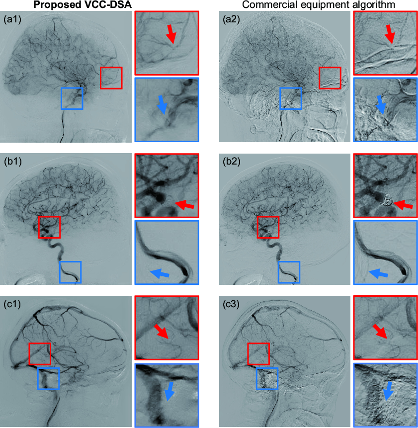
Figure 6: Cross-dataset evaluation—high visual and structural fidelity on a different vendor/device dataset.
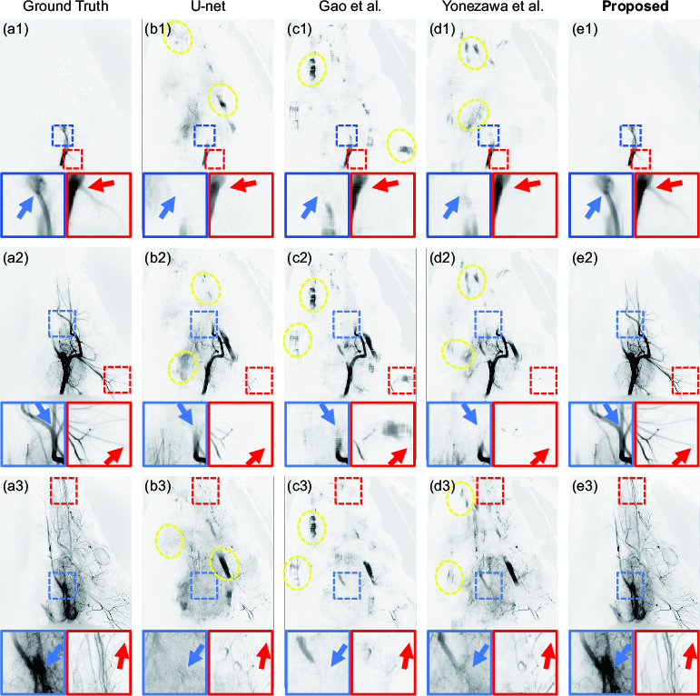
Figure 7: Cross-species generalization—precise vascular imaging and artifact suppression trained on human, tested on animal data.
Ablation Analyses
- LSMP: Excluding mask information severely degrades quantitative performance (SSIM drop from 96.8% to 90.7%, PSNR reduction from 32.6 dB to 23.7 dB), demonstrating its necessity for resolving anatomical ambiguity (Figure 8).
- VCS: Increasing λ directly reduces motion artifact retention, up to an optimal regime, beyond which overregularization can suppress fine vascular features (Figure 9, Figure 10).
- MDSS: Incorporating mixup-based augmentation leads to further quantitative improvements (SSIM from 98.3% to 98.8%, PSNR from 41.3 to 43.2 dB) and reduces misidentification of non-vascular structures as vessels (Figure 11).
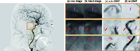
Figure 8: Ablation of LSMP demonstrates a clear performance gap in vessel realism and artifact suppression.
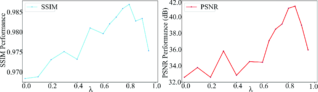
Figure 9: Quantitative effect of VCS scaling: both SSIM and PSNR improve as λ increases until oversmoothing occurs.
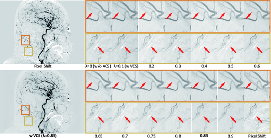
Figure 10: Visual effect of VCS scaling—artifact reduction with increased λ and superior performance over the commercial Pixel Shift method.
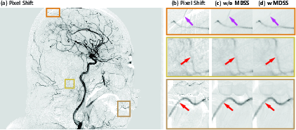
Figure 11: MDSS enhances vessel discrimination, suppresses tube/teeth artifacts, and improves label quality and vascular visibility.
Robustness to Motion Severity
Performance remains high even under simulated large-amplitude motion artifacts; SSIM and PSNR only decrease modestly at the most extreme (>5 SD displacement) levels, underscoring the model's clinical reliability (Figure 12).
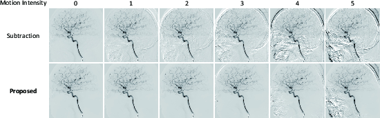
Figure 12: The model preserves vascular clarity and suppresses motion artifacts across the full range of realistic patient movement scenarios.
Failure Analysis
Most failure cases are associated with highly complex and coordinated compound motion (e.g., rapid eyeball rotation affecting adjacent vessels), where intricate dynamic and shape similarity exceed the current model's representational capacity (Figure 13).
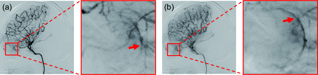
Figure 13: Failure example illustrating the challenge in suppressing motion artifacts that share curvature and intensity with authentic vessels.
Implications and Future Directions
VCC-DSA mitigates fundamental challenges in artifact-prone DSA imaging by combining architectural design, robust self-consistency regularization, and adaptive data/label evolution to minimize reliance on rare high-quality supervised labels. Practically, its adoption can substantially reduce repeat imaging and consequent patient risk in acute neurovascular and endovascular interventions, improve temporal and diagnostic resolution, and increase automation potential in perioperative and intraoperative DSA workflows.
Theoretically, this research contributes to structured learning under noisy supervision in medical imaging, introducing paradigms for exploiting inter-image and label consistency amidst physiologic variability and imperfect gold standards. Extensions to nonlinear motion fields, dynamic three-dimensional reconstructions, and generalized vascular/tissue domain transfer remain promising avenues.
Conclusion
VCC-DSA establishes a robust framework for precise, artifact-suppressed DSA synthesis, validated on both clinical and experimental datasets. Leveraging learning-based subtraction, residual dense architectures, vascular consistency constraints, and self-evolving label strategies, it achieves strong quantitative and qualitative gains and robust cross-domain generalization. This work evidences the impact of architectural and loss-function-level domain knowledge integration in addressing deep learning ill-posedness and practical clinical constraints in interventional imaging.
Reference: "VCC-DSA: A Novel Vascular Consistency Constrained DSA Imaging Model for Motion Artifact Suppression" (2604.10700)