- The paper introduces a masked loss function leveraging automated organ masks to enforce anatomical plausibility in AAA segmentation.
- The method, built on a 2D U-Net, achieves a Dice coefficient increase from 0.637 to 0.914 with limited training data.
- The approach reduces false positives from adjacent organs, enabling precise 3D morphological analysis for clinical intervention planning.
Introduction
Accurate segmentation of abdominal aortic aneurysms (AAAs) in computed tomography angiography (CTA) is a foundational step in vascular diagnosis and intervention planning. The inherent anatomical complexity, low-contrast vessel boundaries, and proximity of visually similar organs substantially degrade conventional CNN-based segmentation performance. The paper "Anatomy-Informed Deep Learning for Abdominal Aortic Aneurysm Segmentation" (2604.10312) addresses these challenges by integrating explicit anatomical priors—derived from automatically generated organ masks—into the deep learning pipeline for AAA segmentation. This approach aims to suppress anatomically implausible predictions, enhance boundary consistency, and boost robustness, particularly in limited-data regimes.
Methodology
The proposed method employs TotalSegmentator to generate exclusion masks for non-vascular organs from CTA volumes. These masks are integrated into a masked loss function, which restricts optimization to anatomically plausible aortic regions, thereby regularizing the learning process. A standard 2D U-Net serves as the backbone, trained using a weighted sum of masked Dice and binary cross-entropy losses, with pixels from excluded regions omitted from gradient updates. The exclusion masks are derived automatically, avoiding any additional manual annotation or architectural modifications.
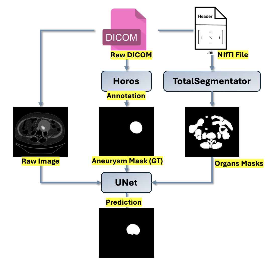
Figure 1: Pipeline of the proposed method from preprocessing to training.
The dataset comprises 20 expertly annotated patient CTA scans, preprocessed to isotropic 1.0mm3 voxels with strong data augmentation. Training, validation, and test splits are strictly at the patient level to preclude data leakage. Extensive augmentation (rotations, elastic deformations, intensity shifts) counteracts overfitting due to the limited training examples.
Quantitative and Qualitative Analysis
The anatomy-aware U-Net achieves a significant increase in segmentation accuracy over the baseline. For a regime with 10 training patients, the mean Dice coefficient improves from 0.637 (baseline) to 0.914 (anatomy-aware). When scaling to 16 training patients, the mean Dice stabilizes at 0.906, confirming the generalization benefit. Both recall and precision are substantially increased, and the standard deviation in performance narrows, indicating improved consistency. The improvement is particularly notable in anatomically complex cases where bowel, bone, or paraspinal musculature abuts the aneurysm, a dominant source of false positives for intensity-based deep models.
Qualitatively, the anatomy-informed model eliminates most off-aorta activations, yielding segmentations closely conforming to ground-truth annotations and preserving smooth, realistic boundaries. Visualizations highlight the marked reduction in erroneous labeling—critical for downstream morphometric analyses.
3D Reconstruction and Morphometric Analysis
3D reconstruction of the aneurysm surface from 2D segmentation masks enables extraction of clinically pertinent shape and size metrics: maximal diameter, surface area, and total aneurysm volume. The anatomy-aware model's segmentations result in surface and volumetric measures that closely approximate ground-truth values, generally within a clinically acceptable margin.
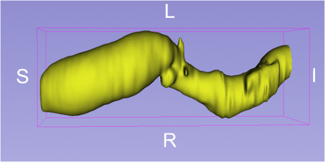
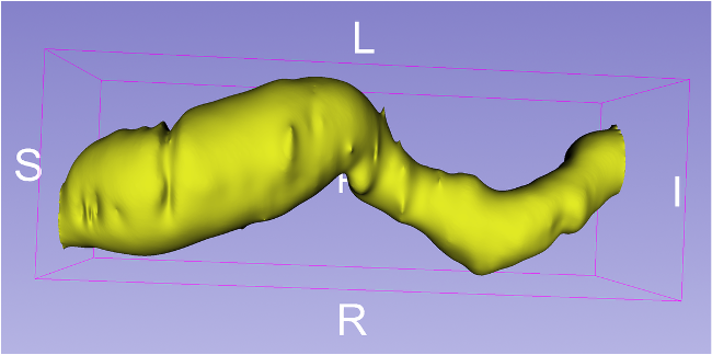
Figure 2: 3D reconstructed aneurysm from predicted segmentation (right) and corresponding ground truth (left).
Advanced morphometric descriptors, such as centerline extraction, diameter variation, and local curvature, become reliable when false positives are suppressed and segmentation matches anatomic boundaries. These descriptors are critical for biomechanics-based rupture risk assessment and for planning of personalized vascular interventions.
Discussion and Implications
The explicit use of organ exclusion masks fundamentally regularizes the segmentation problem, introducing anatomical constraints that are agnostic to intensity distribution but tightly coupled to plausible vascular anatomy. This suppresses improbable activations in regions such as bowel or bone, overcoming one of the principal failure modes of traditional U-Nets in low-contrast, multi-structure imagery. The marked segmentation performance gains are achieved without complex architectural modification or manual annotation of additional structures, retaining methodological simplicity and computational efficiency.
This approach is particularly advantageous in the context of limited and imbalanced medical datasets—a persistent constraint in vascular imaging—since exclusion masks amplify signal quality and decrease the need for extensive manual curation. The framework is broadly extensible to other vascular and organ segmentation tasks where anatomical context is fundamental.
Future work could leverage 3D segmentation models to further capitalize on spatial context, or introduce soft rather than hard exclusion masks to permit more nuanced anatomical regularization. Integration with semi-supervised or unsupervised learning paradigms could be particularly synergistic in even more data-scarce environments.
Conclusion
This paper demonstrates that anatomy-informed loss masking, using fully automated organ segmentation, markedly improves AAA segmentation in challenging CTA data, producing reliable and anatomically plausible results even when annotated data are scarce. The methodology is simple, requires no architectural change, and is readily transferable to other clinically significant segmentation tasks in medical imaging. This represents a substantive advance in anatomically-regularized deep learning for medical image analysis.