- The paper demonstrates that realistic degradation simulation coupled with latent diffusion can elevate EICT scans to PCCT-like quality.
- It employs a continual-learning autoencoder with clinically validated artifact simulation to achieve up to 35.9% SSIM and 19.5% PSNR improvements.
- Enhanced scans improve downstream clinical metrics, boosting detection sensitivity, F1 scores, and AUC in multi-center datasets.
Distilling PCCT Quality into Routine Chest CT via Clinically Validated Degradation Modeling
Introduction
Photon-counting CT (PCCT) has emerged as a significant advance in medical imaging by delivering higher spatial resolution and reduced noise relative to conventional energy-integrating CT (EICT) systems. However, due to constraints in cost and limited hardware penetration, PCCT remains inaccessible for large-scale clinical and research applications. The present study introduces a novel, clinically validated degradation-to-enhancement framework for distilling the benefits of PCCT into routine EICT. The approach is centered on simulating realistic EICT acquisition degradation from PCCT data and then training a deep learning model to invert this process, thereby enhancing EICT scans to approximate PCCT-like quality.
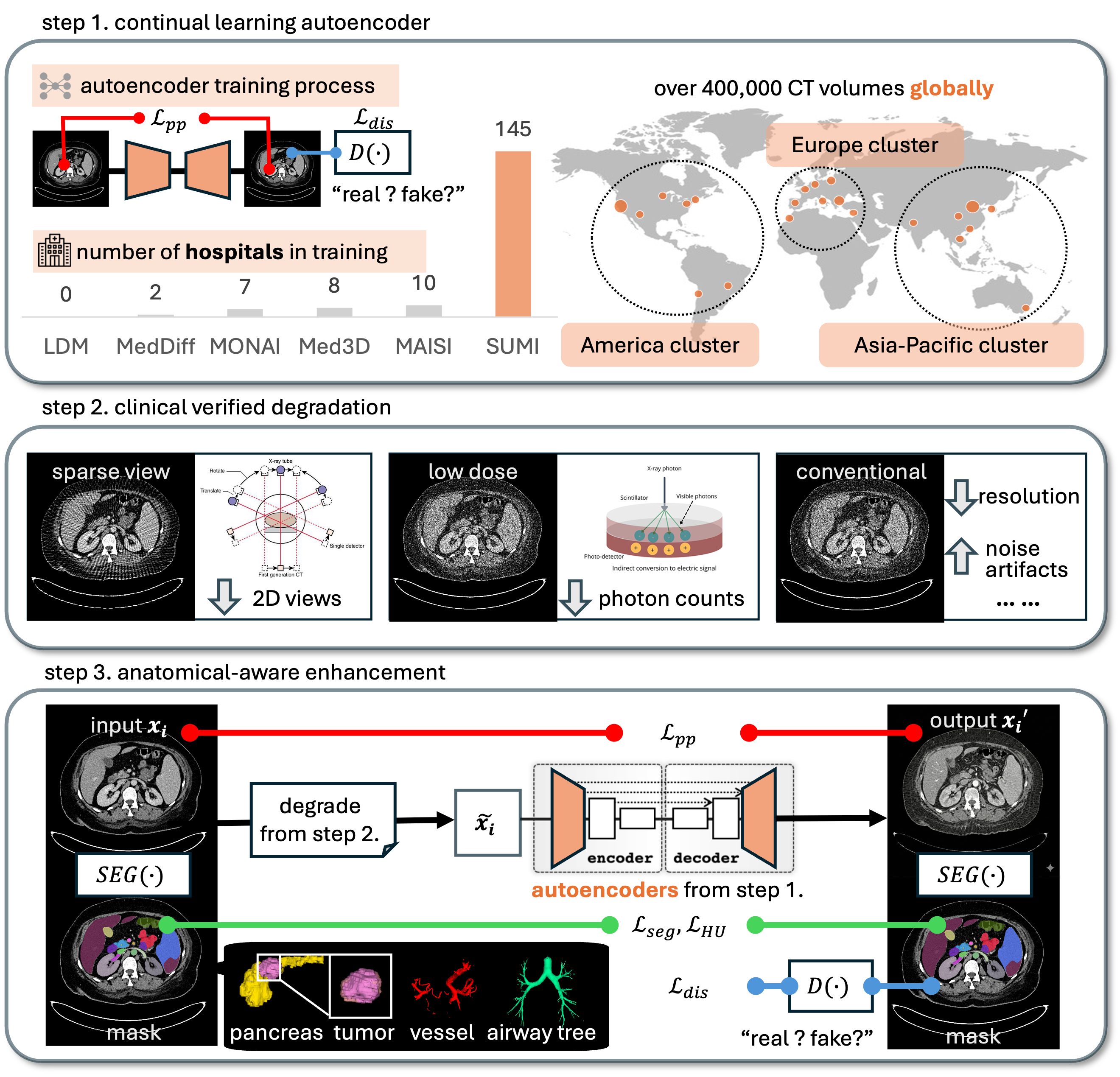
Figure 1: Overview of the proposed framework, including large-scale autoencoder pretraining, clinically validated degradation simulation, and reverse enhancement via a diffusion model.
Methodology
Continual Learning Autoencoder
The model begins with a continual-learning autoencoder pretrained on an extensive and diverse dataset of over 405,000 EICT scans and 1,046 PCCT scans sourced from 145 hospitals across 19 countries. This architecture incorporates a memory loss mechanism, supporting robust anatomical representation learning, domain generalization, and site-specific adaptability. The large-scale pretraining serves as a backbone for downstream generative tasks in medical imaging.
Clinically Validated Degradation Simulator
A key contribution is the development and clinical validation of a degradation simulator. The simulator degrades high-quality PCCT scans via three major modalities, each corresponding to authentic EICT artifacts:
- Sparse-View Degradation: Reduces the number of acquisition projections, mimicking streak artifacts.
- Low-Dose Simulation: Models dose-reduction protocols by injecting photon-statistics-driven Poisson noise.
- Conventional Degradation: Incorporates spatial downsampling and mixed electronic noise, replicating standard EICT characteristics.
Expert radiologists confirm the realism of synthesized degradations, ensuring clinically plausible training data without requiring paired PCCT/EICT acquisitions.
Enhancement via Latent Diffusion
Training proceeds in the latent space of the pre-trained autoencoder. The model employs a latent diffusion framework that learns to enhance degraded PCCT or EICT images back toward reference PCCT quality. The training objective integrates four loss components:
Experimental Evaluation
Data and Baseline Comparison
Evaluation leverages a multi-faceted dataset, including 405,379 EICT scans for pretraining, 1,046 PCCTs for enhancement training, and a curated set of 17,316 public EICT scans enhanced to PCCT-quality with comprehensive voxel-wise anatomical annotations. Comparisons are performed against state-of-the-art (SOTA) restoration methods from filtering (NLM), transformer-based (Swin2SR), GAN (Pix2Pix, SRGAN), and diffusion (SR3, NEED) domains.
Image Quality and Clinical Metrics
The proposed method consistently outperforms baselines, achieving up to 35.9% improvement in SSIM and 19.5% in PSNR across external patient datasets in all tested degradation modalities. Ablation analysis confirms that each simulated degradation form is critical to robust real-world generalization.
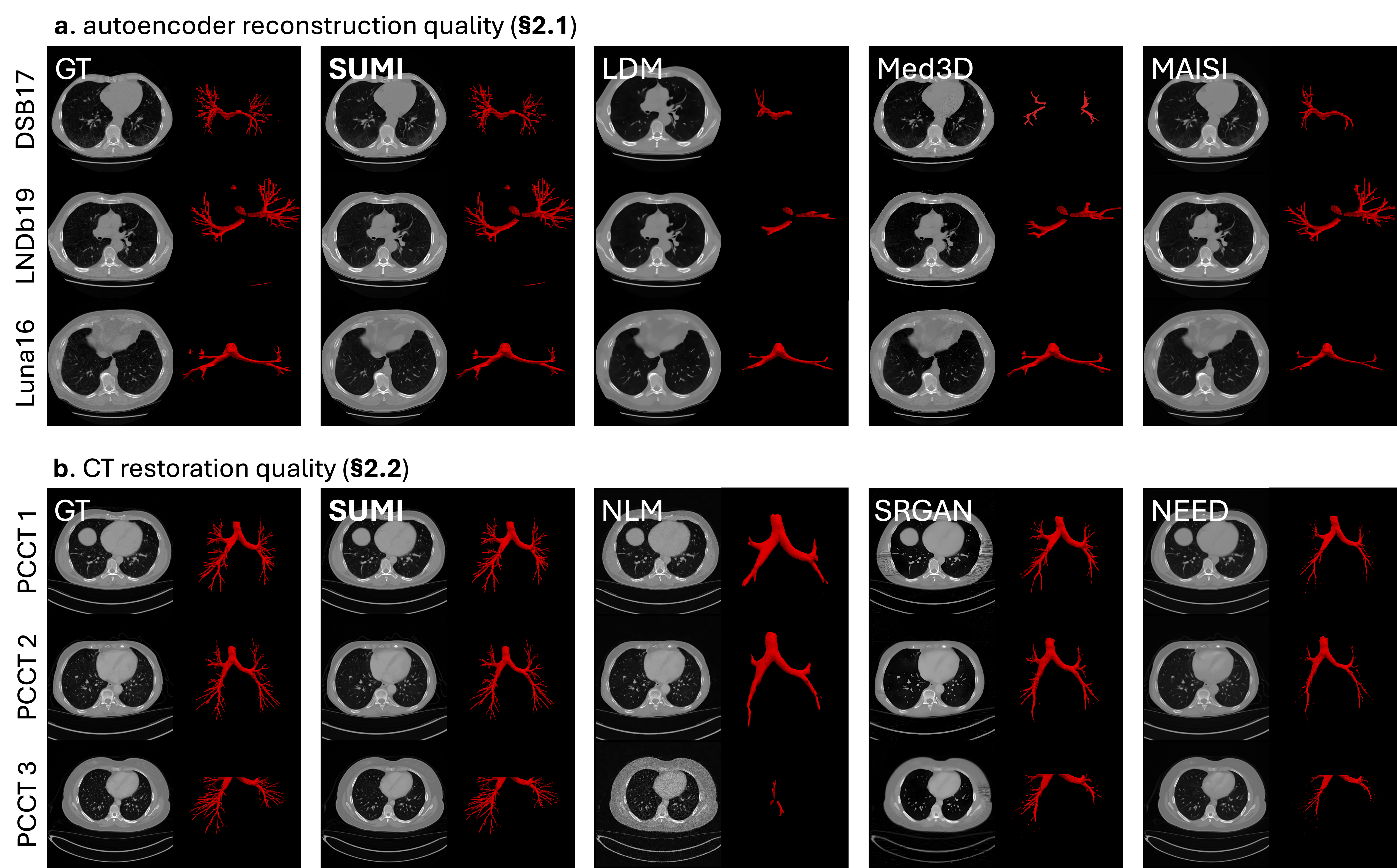
Figure 3: Cross-dataset visual evaluation shows superior generalization, notably preserving fine airway topologies under domain shift. PCCT enhancement demonstrates greater fidelity to ground truth anatomical features compared to strong baselines.
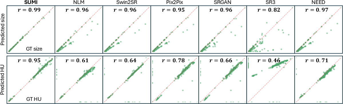
Clinical evaluations utilize Pearson correlation of organ-specific HUs and segmentation masks, confirming that the model retains structural accuracy without synthesizing hallucinated features. Integration of enhanced scans into SOTA detection pipelines yields downstream sensitivity and F1 improvements of up to 15.2%, with AUC gains of 10.5% in multicenter test sets.
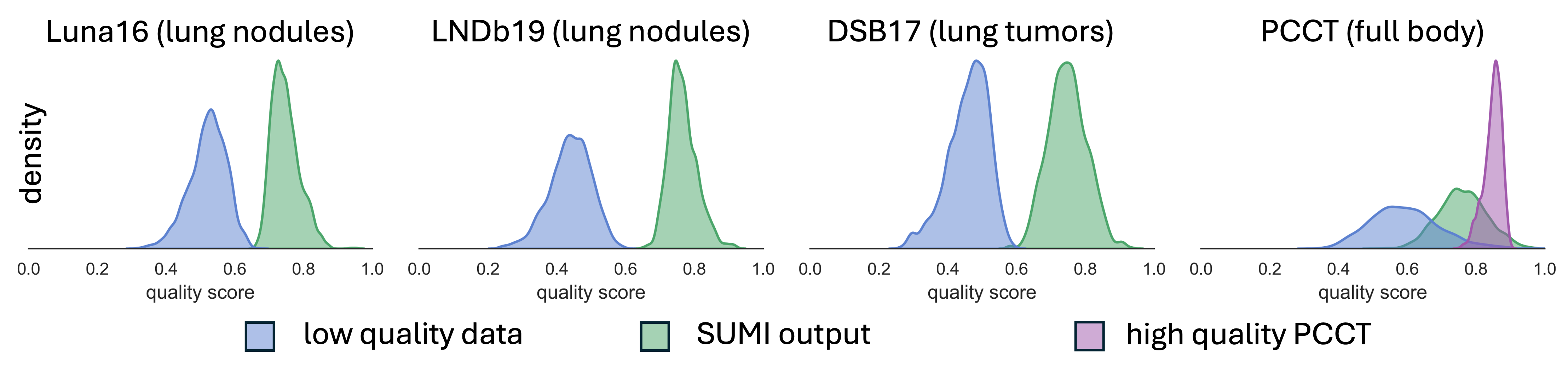
Figure 4: The enhancement process shifts quality score distributions on public chest CT datasets toward the PCCT standard, indicating robust and consistent quality improvements.
Implications and Future Directions
The methodological framework demonstrates that it is possible to systematically translate the gains of advanced imaging hardware into legacy systems using principled, reversible degradation simulation and controlled enhancement via generative models. Practically, this enables broader, more equitable access to high-fidelity imaging for both clinical deployment and research applications, circumventing the high costs and limited access associated with PCCT hardware.
Theoretically, the study underscores the importance of domain-aware degradation modeling and thorough clinical validation in clinical AI development. The use of a continual-learning autoencoder backbone establishes a scalable foundation for multi-institutional, multi-domain generalizability and for extensibility to new generative medical imaging tasks.
Future work should address extension beyond the thoracic region and progression from 2D to 3D volumetric enhancement to further optimize z-axis continuity. Prospects include exploring fully unsupervised domain adaptation, integrating spectral information, and extending enhancement paradigms to other imaging modalities.
Conclusion
This work presents a scalable, clinically validated strategy for distilling the benefits of photon-counting CT into routine EICT via realistic degradation simulation and deep generative enhancement. The framework achieves strong quantitative and qualitative gains in image quality, anatomical fidelity, and downstream clinical detection across diverse and externally validated chest CT datasets. These findings support the systematic transfer of emerging imaging advances to standard clinical workflows, fostering reproducible research and improved healthcare outcomes.