- The paper introduces a novel geometric framework that learns an energy landscape over multi-sequence MRI data to serve as a label-free baseline for tracking tissue states.
- It employs denoising score matching within a compact implicit neural representation to map voxel intensity vectors, distinguishing healthy and tumor regimes via stable energy basins.
- Longitudinal analysis reveals measurable energy shifts that correlate with tissue transitions, enabling early detection of tumor recurrence before visible anatomical changes.
Energy-Based Tissue Manifolds for Longitudinal Multiparametric MRI Analysis
The paper "Energy-based Tissue Manifolds for Longitudinal Multiparametric MRI Analysis" (2604.07180) introduces a geometric paradigm for patient-specific longitudinal mpMRI analysis by learning an energy landscape directly over MRI sequence space. Unlike conventional spatial or segmentation-based methods, each voxel is mapped to a multi-sequence intensity vector (T1,T1c,T2,FLAIR,ADC) in Rd, capturing multidimensional tissue composition. A compact implicit neural representation (INR) models a scalar energy function Eθ(u) via denoising score matching, efficiently trained on the baseline scan without reliance on segmentation masks or spatial coordinates.
Basins in the energy landscape correspond to stable tissue regimes, with minima representing healthy and tumour states. The gradient and curvature of Eθ encode proximity to boundaries and local tissue constraints, respectively. The baseline energy manifold, defined by patient-specific acquisition protocol, serves as a geometric reference for all subsequent scans. Longitudinal analysis is reframed as geometric evaluation: monitoring how follow-up sequence vectors evolve relative to the fixed baseline energy landscape, rather than comparing evolving anatomical masks.
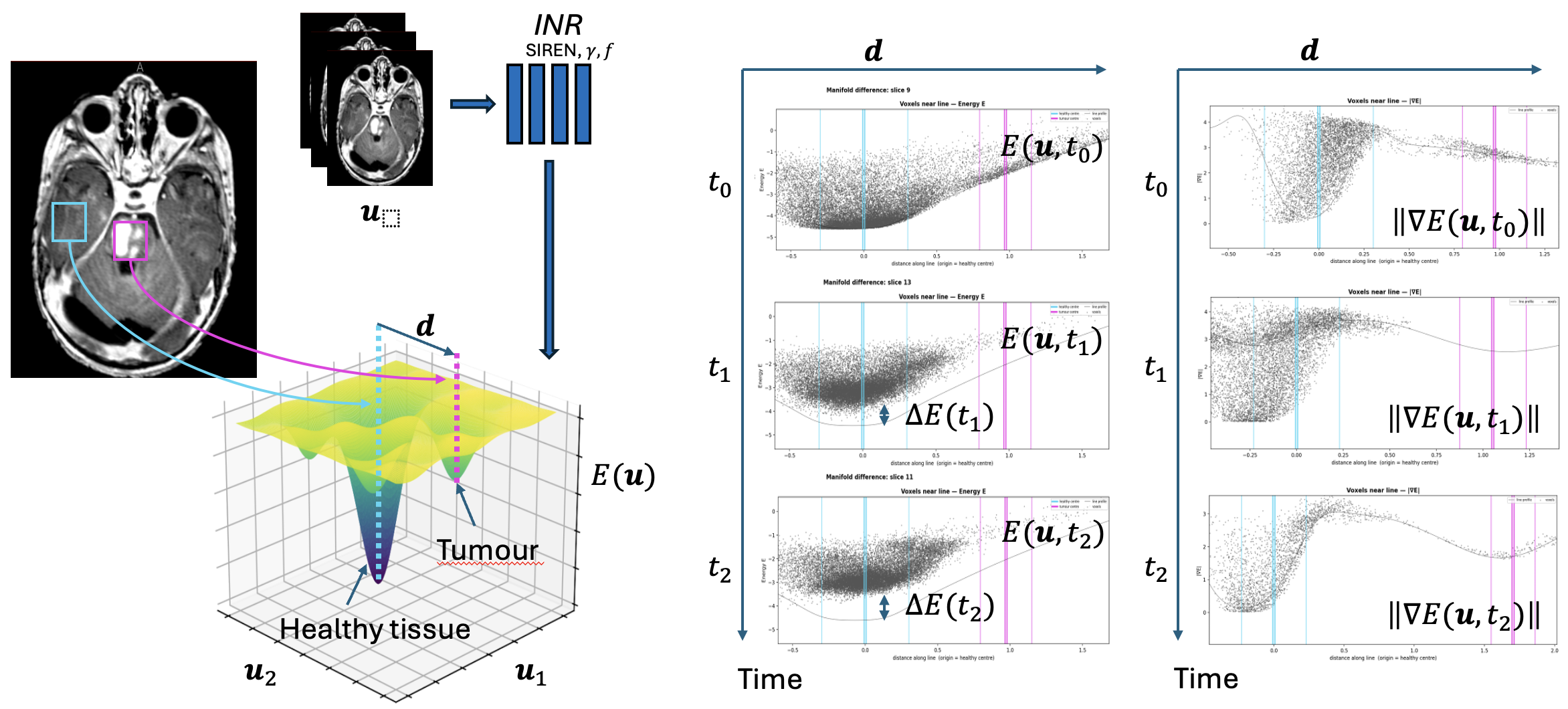
Figure 1: Overview of the proposed energy-based longitudinal tissue tracking framework, showing ROIs in baseline scan defining attractors in sequence space and the healthy--tumour axis for longitudinal projection and analysis.
Score-Based Model Training and Architectural Details
Energy function modeling employs denoising score matching (DSM), circumventing partition function computation and enabling efficient estimation of the data score in low-dimensional sequence space (d=5). The INR employs sinusoidal activations (SIREN) and Gaussian Fourier feature encodings, benefiting from smooth differentiability required for both score estimation and geometric analysis. Compact architecture and single noise scale are sufficient due to the natural contrast clustering in the MRI modalities considered. Training completes in minutes per patient, facilitating rapid manifold creation.
This approach differs from diffusion model frameworks commonly seen in recent medical imaging literature [ho2020denoising; yang2023diffusion], which operate in image space for synthesis, reconstruction, or anomaly detection [wolleb2022diffusion; liu2025longitudinal]. Here, generative objectives are leveraged purely for geometric reference construction—not for image generation—and operating directly in sequence space provides tighter coupling to tissue biophysics.
Geometric Analysis of Tissue Regimes
The learned Eθ produces a stratified tissue manifold where basin attractors are manually anchored at healthy and tumour ROIs in the baseline scan. The healthy--tumour centroid axis in sequence space provides a one-dimensional coordinate for tracking regime evolution. The differential geometry—basin width, energy barrier height, gradient magnitude, and Laplacian curvature—allows precise quantification of tissue stability, inter-regime transition smoothness, and local risk structure.
Longitudinal scans are projected into the fixed baseline energy manifold without retraining, permitting rigorous, label-free geometric comparison. This is fundamentally distinct from methods reliant on longitudinal anatomical segmentations, which suffer from unstable or ill-defined boundaries, particularly early in recurrence.
Case Studies: Stable Disease vs. Recurrence
Two paediatric brain tumour cases illustrate the framework's interpretive power:
- Stable Disease: Post-resection scans over two years exhibited minimal sequence-space drift, with healthy tissue vectors remaining confined to the original low-energy basin and no systematic shift toward the tumour regime. Energy barrier and basin width remained stable.
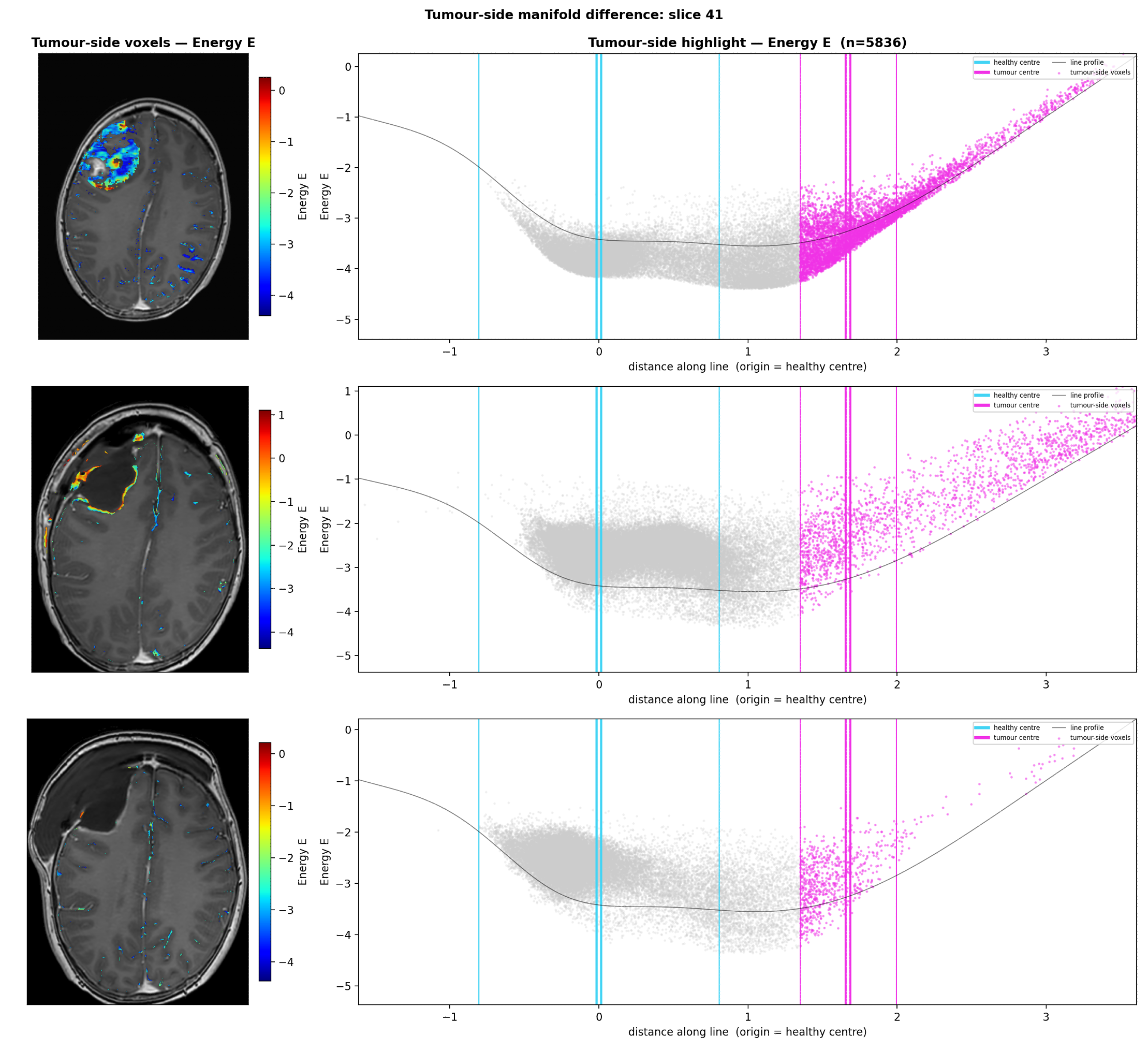
Figure 2: Longitudinal projection of voxel energies for a patient with stable disease, showing preserved basin structure and absence of regime shift.
- Recurrence: In the recurrence case, pre-anatomical tumour resurgence was detected as progressive displacement of voxel vectors toward the original tumour basin in sequence space, accompanied by increased mean energy levels and deformation of the energy profile. At radiological confirmation, a new tumour basin emerged, indicating that sequence-space energy drift can precede visible anatomical manifestation.
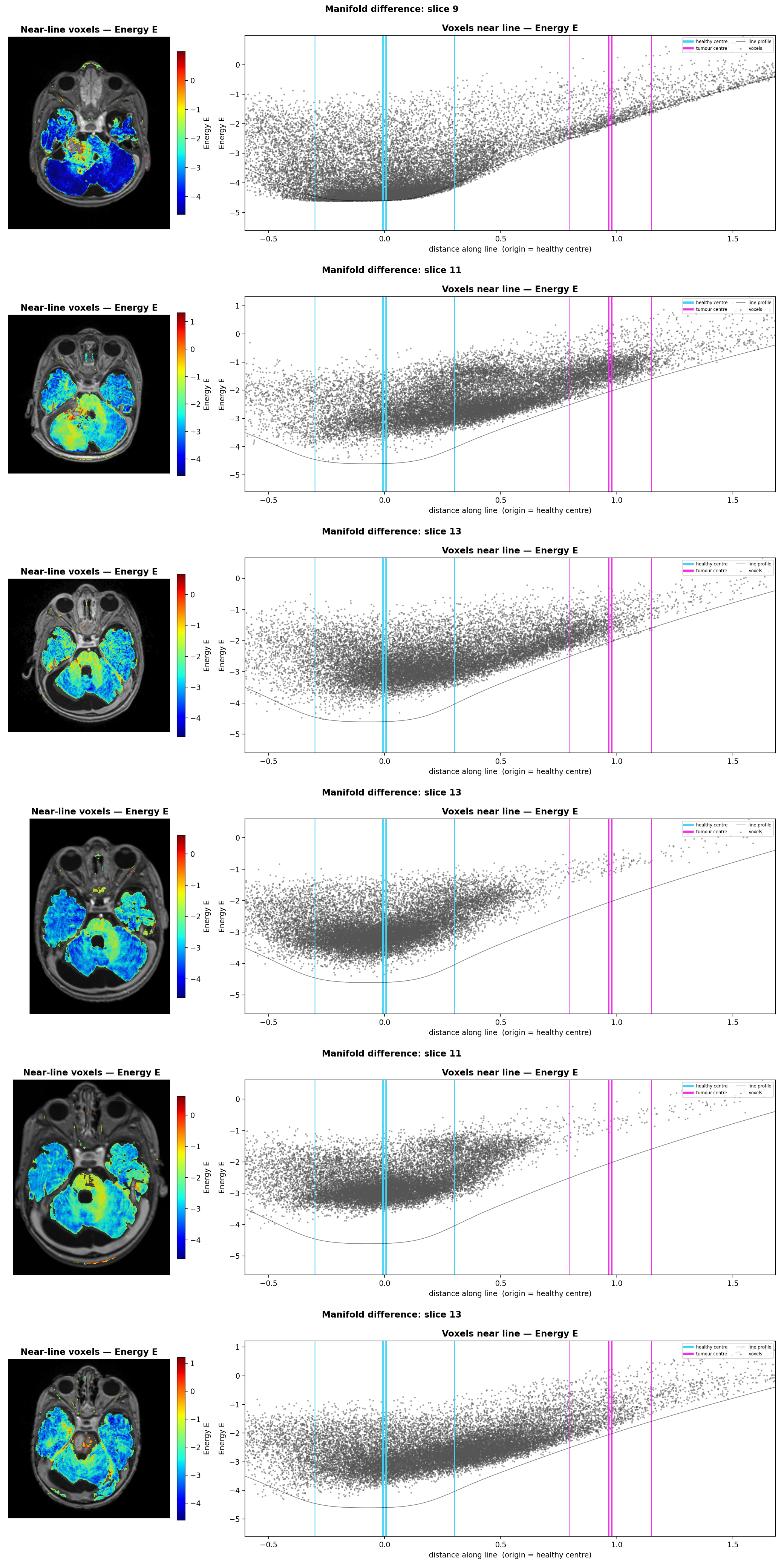
Figure 3: Longitudinal projection of voxel energies for a patient with recurrence, demonstrating displacement toward tumour regime and emergence of a new sequence-space basin.
Strong numerical results corroborate the power of geometric tracking: Mean energy change (δE) and sequence-space drift for recurrence are consistently positive and statistically significant, while stable disease exhibits negligible or negative drift.
Practical and Theoretical Implications
This geometric approach establishes a rigorous, coordinate-independent representation for patient-specific longitudinal analysis in oncology, free from segmentation boundary dependency. It fundamentally shifts the paradigm from spatial mask evolution to geometric deviation, suggesting that tissue at risk can be detected as sequence-space drift prior to anatomical recurrence.
The implications extend to early intervention, post-treatment monitoring, and cross-patient stratification, with potential for integration into population-level analysis of tissue regime geometries. The framework's robustness, sensitivity to early relapse, and scalability need comprehensive validation across larger cohorts and disease types. Incorporation of spatial priors and extension to more modalities could further enhance tracking accuracy and interpretability.
Conclusion
The energy-based tissue manifold methodology constitutes an authoritative geometric foundation for label-free, patient-specific longitudinal mpMRI analysis. By leveraging sequence-space modeling and denoising score-based learning, it enables interpretable regime tracking, early risk detection, and a rigorous reference system for longitudinal neuro-oncology studies. Future developments may include multi-modal integration, automated ROI anchoring, and population-level comparative manifold analysis, with potential translational impact for early relapse detection and personalized monitoring in the clinical workflow.

