- The paper introduces an infrared tomography lab that minimizes uncertainty to below 25 mK for detecting subtle sub-0.1 K temperature variations.
- It employs rigorous calibration protocols including drift correction, spatial normalization, and stable mechanical alignment to enhance measurement precision.
- The system’s performance, validated via phantom studies and 3D reconstructions, supports future AI-driven clinical imaging applications.
Introduction and Rationale
This paper establishes a rigorous experimental protocol and laboratory platform enabling steady-state infrared (IR) tomography for medical imaging applications, with a focus on resolving sub-0.1 K surface temperature perturbations induced by modest internal contrasts (ΔT=1–3 K) at depths consistent with biological tissue. The work directly addresses a historical deficiency in IR-based medical diagnostics—the lack of reproducible, quantitatively characterized data acquisition under tightly controlled environmental and instrumental conditions.
Current advances in IR tomography are predominantly algorithmic and computational, but their efficacy is fundamentally limited by measurement stability and calibration rigor. The methodology articulated here explicitly targets minimization of instrumental drift, suppression of environmental fluctuations, and robust system-level calibration as prerequisites to quantitative tomographic inversion.
System Requirements and Design Criteria
The laboratory platform was developed to resolve weak surface temperature variations driven by internal thermal heterogeneities transported via diffusion—not direct radiative emission. This places stringent demands on measurement uncertainty, boundary-condition stability, and geometric registration. The paper specifies operational performance thresholds:
- Relative temperature resolution ≤ 25 mK
- Operational precision (environmental + instrumental) ≤ 50 mK
- Spatial non-uniformity (corrected) ≤ 30 mK
- Frame-to-frame drift (30 min cycle) ≤ 30 mK
Mechanical alignment criteria and phantom design guidelines (including conductivity, internal referencing, and geometric reproducibility) are also delineated. The dominance of system-level perturbations (convective, radiative, conductive, and mechanical) over detector-limited noise is emphasized as the principal metrological constraint.
Laboratory Implementation
The experimental assembly consists of a carefully engineered thermal enclosure constructed from acrylic, with airflow suppression and radiative background stabilization via a water-filled container positioned behind the phantom.
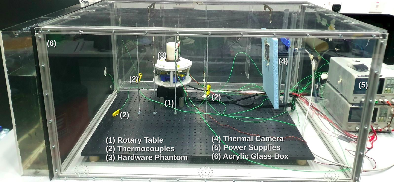
Figure 1: The acrylic enclosure stabilizes local thermal conditions, suppresses convective currents, and isolates the phantom from laboratory variations.
Thermal acquisition utilizes a FLIR A400 IR camera with a sub-40 mK NETD, equipped with in-field reference resistors and a Π-shaped high-emissivity calibration surface.
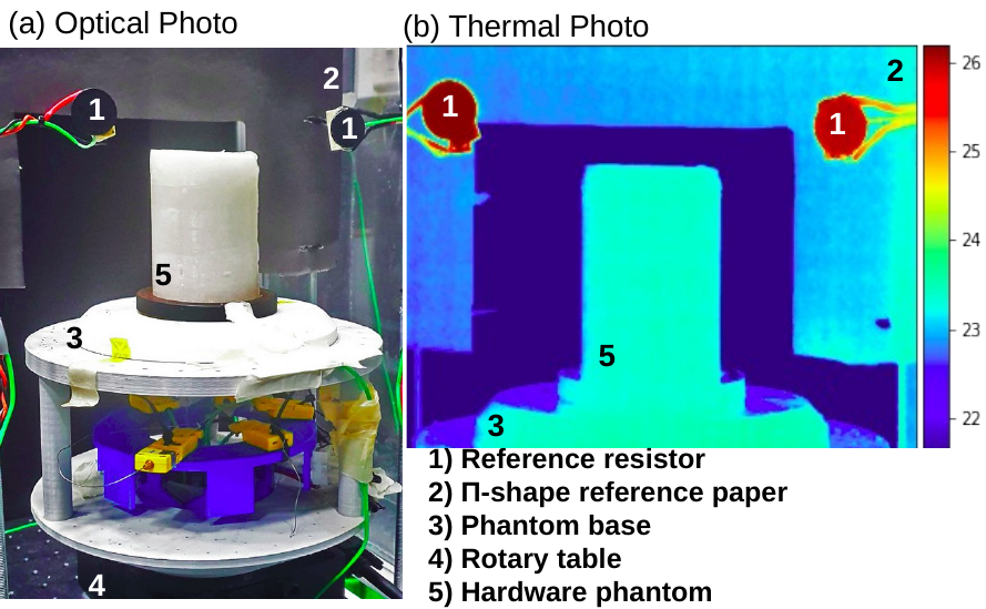
Figure 2: Optical and thermal registration of reference resistors and the Π-surface enables robust spatial and temporal correction.
Mechanical positioning is achieved with a motorized rotary stage, maintaining sub-0.5 mm centering and 0.02° step reproducibility, essential for tomographic angular sampling. All major heat-generating electronics are thermally decoupled from the measurement volume.
Acquisition is automated (LabVIEW-based), and the protocol uses interleaved projection sequences to decouple systematic drift and projection angle. Simultaneous temperature readings from embedded thermocouples, air, and references accompany each thermal frame.
Calibration Pipeline and Correction Methodology
Quantitative inversion under the described model necessitates absolute minimization and correction of additive and multiplicative errors:
- Thermocouple Offset Alignment: All internal references are cross-calibrated, ensuring offset consistency.
- Temporal Drift Correction: Frame-by-frame drift is suppressed using embedded resistive references as in-field anchors.
- Environmental Drift Alignment: Slow air temperature drift is fit and subtracted over the full acquisition interval.
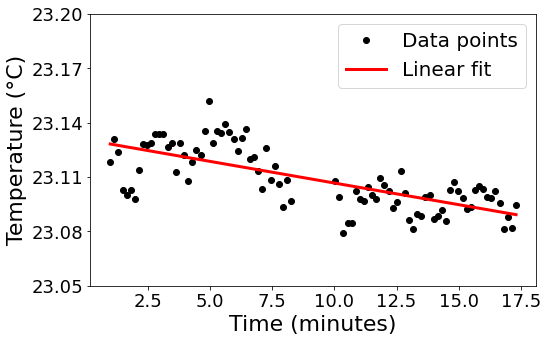
Figure 3: Air temperature within the enclosure is characterized and corrected using linear drift modeling across the acquisition window.
- Spatial Non-Uniformity Correction: The Π-shaped isothermal surface enables columnar and row-wise normalization, suppressing fixed-pattern artifacts and pixel response heterogeneities.
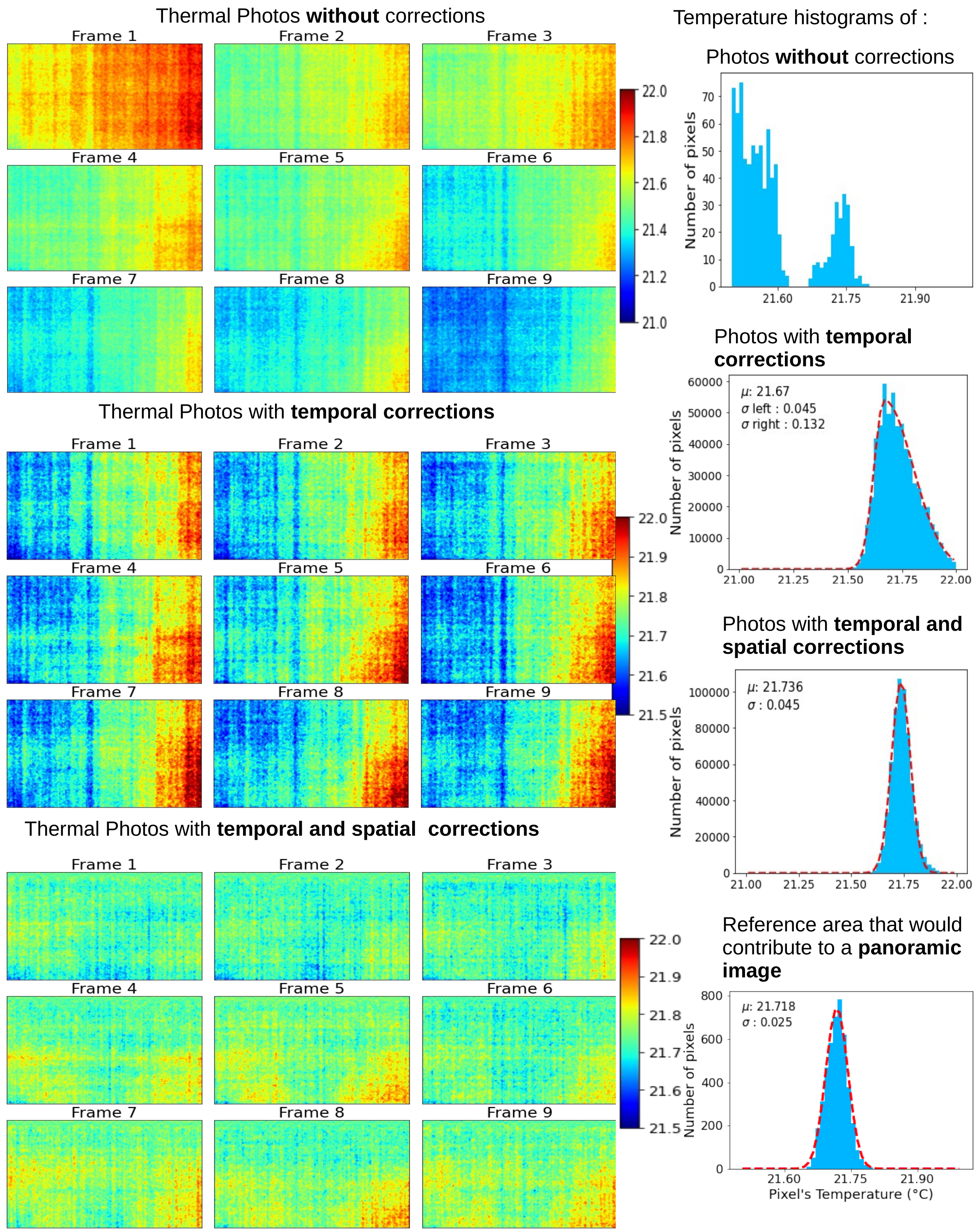
Figure 4: Correction pipeline output—top: raw; middle: temporally corrected; bottom: temporally and spatially corrected images and corresponding histograms. Final uncertainty is reduced to ∼25 mK.
The calibrated dataset is then restructured into panoramic projections by concatenating narrow frontal surface strips at each angle, yielding input amenable to tomographic inversion.
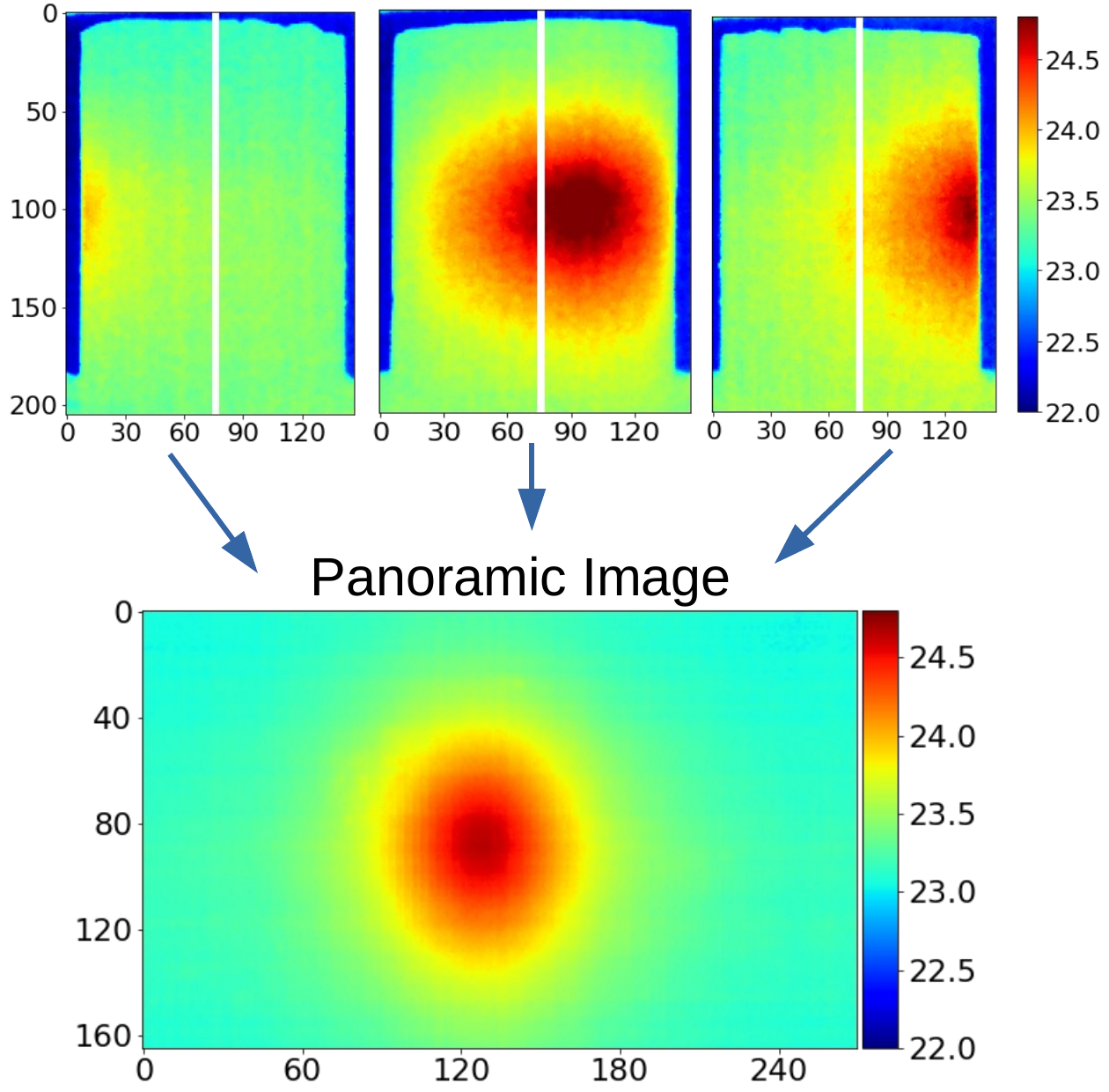
Figure 5: Construction of panoramic projection data combines angularly sampled surface temperature strips into a consistent tomographic dataset.
The laboratory achieves ≤25 mK global uncertainty in panoramic projection data after full correction, validated both statistically and via calibration standards.

Figure 6: Frame averaging suppresses stochastic detector noise, contributing to millikelvin-level precision.
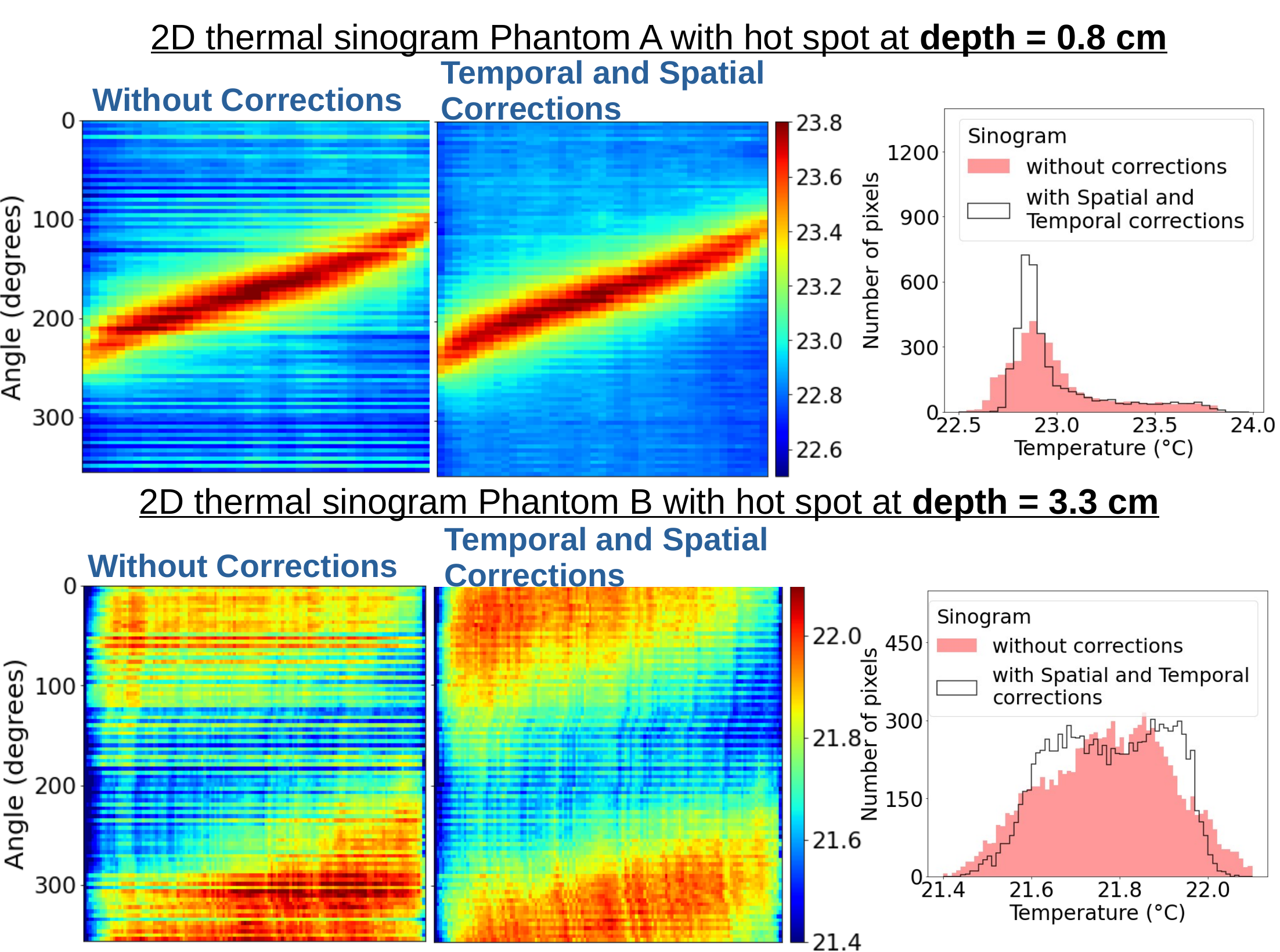
Figure 7: Correction impact on sinograms from phantoms with embedded heat sources—background fluctuation is suppressed, and source signal becomes separable.
Reconstruction robustness is benchmarked via phantoms with embedded controlled heat sources and independent thermocouple readouts. The AMIAS/RISE inversion framework accurately localizes and quantifies internal temperature distributions, with reconstructed values in direct agreement (<0.15 K discrepancy) versus internal probe measurements and micro-CT for source position validation.

Figure 8: 3D reconstructed temperature (AMIAS/RISE framework) for a dual-source phantom alongside μCT anatomical reference—demonstrating topological and quantitative concordance.
The main sources of measurement uncertainty—detector drift, spatial non-uniformity, and environmental fluctuation—are all quantified with and without mitigation, with tabulated reductions supporting the achieved performance claims.
Implications and Future Developments
The laboratory protocol detailed here establishes the prerequisite metrological infrastructure to support systematic studies in IR tomographic imaging of biological and tissue-analog phantoms. Performance is achieved at a level compatible with detection of physiological-scale contrasts at clinically relevant depths, and the platform directly supports future research in:
- Benchmarking and optimization of physics-guided and AI-driven inversion schemes
- Controlled validation of system performance with diverse phantoms
- Extension to in-vivo studies, contingent on improved environmental isolation and physiological motion correction
- Development of standard calibration procedures and uncertainty quantification metrics for IR-based medical imaging
Given the ill-posed nature of the steady-state diffusive inverse problem, the demonstrated system calibration and measurement repeatability are essential foundations for algorithmic advancement and clinical translation.
Conclusion
This work presents a comprehensive, experimentally validated protocol and laboratory architecture for steady-state infrared tomography targeting medical imaging applications. By integrating environmental stabilization, robust referencing, automated angular acquisition, and systematic calibration, the effective system uncertainty is reduced below 25 mK—sufficient for resolving clinically relevant temperature perturbations at biologically realistic depths. This enables quantitative tomographic inversion validated against independent internal measurements. The established framework provides a rigorous experimental basis for ongoing development of IR tomographic methodologies.

