- The paper presents a fully open-source device that overcomes ergonomic and refractive limitations in MRI by enabling accurate, comfortable visual stimulus presentation.
- It details a modular design with independently adjustable lens holders for precise interpupillary distance correction and rapid tool-less lens switching.
- Practical fabrication using 3D-printable, MRI-compatible materials demonstrates improved participant comfort and enhanced data fidelity in fMRI experiments.
Head Coil-Mounted Vision Correction Device for Magnetic Resonance Imaging
Introduction
The paper "Head Coil-Mounted Vision Correction Device for Magnetic Resonance Imaging" (2604.04966) presents a fully open-source, 3D-printable device designed to enhance the quality and comfort of fMRI experiments by providing ergonomic, cost-effective, and flexible vision correction. Visual stimulus protocols in MRI are widely used, yet a significant proportion of the population requires refractive correction, and existing solutions for MRI present multiple limitations, particularly in the context of inter-pupillary distance (IPD) adjustment, lens switching, and compatibility with tightly-fitting head coils.
Context and Limitations of Previous Designs
Commercial approaches to MRI-compatible vision correction fall into head-mounted systems (goggles) and coil-mounted devices. Head-mounted systems induce ergonomic issues, including facial discomfort and pressure points, especially during extended imaging sessions and for participants with large head sizes. Most coil-mounted solutions do not accommodate individual variability in IPD, thus limiting visual correction accuracy. The inability to quickly switch lenses and adapt to different lens geometries further constrains research flexibility. No commercial hardware supports correction for cylindrical errors (astigmatism) or higher-order aberrations.
Design and Mechanical Architecture
The authors introduce a device manufactured with MRI-compatible, inexpensive plastics (notably PLA), fabricated via filament deposition modeling (FDM) and designed in SolidWorks. The device is composed of several modules: a robust head coil mount, interchangeable and independently adjustable lens holders, and non-metallic fasteners. The engineering process included reverse engineering the geometry of the commercially prevalent Siemens 32-channel head coil, using reference photography and SolidWorks’s Sketch Picture Tool to align mechanical CAD models with the coil anatomy.
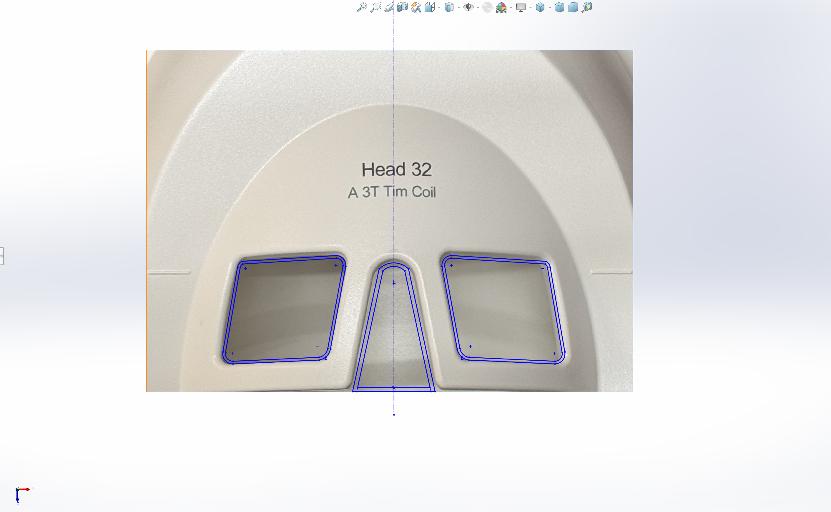
Figure 1: The workflow of adapting the anterior head coil geometry using the Sketch Picture tool in SolidWorks for mount design.
The lens holders exhibit independent horizontal translation, thus offering full IPD adjustment within the constraints of the coil geometry and lens form factor. Modular lens holder assemblies accommodate a wide range of available spherical correction lenses. The fastening system is compatible with both printed and commercial nylon hardware, supporting complete elimination of metal from the assembly.
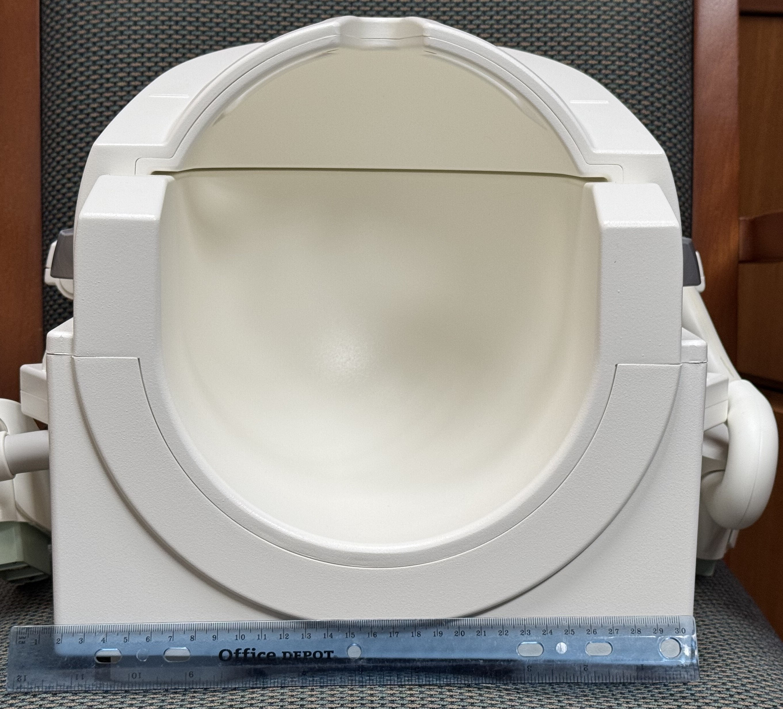
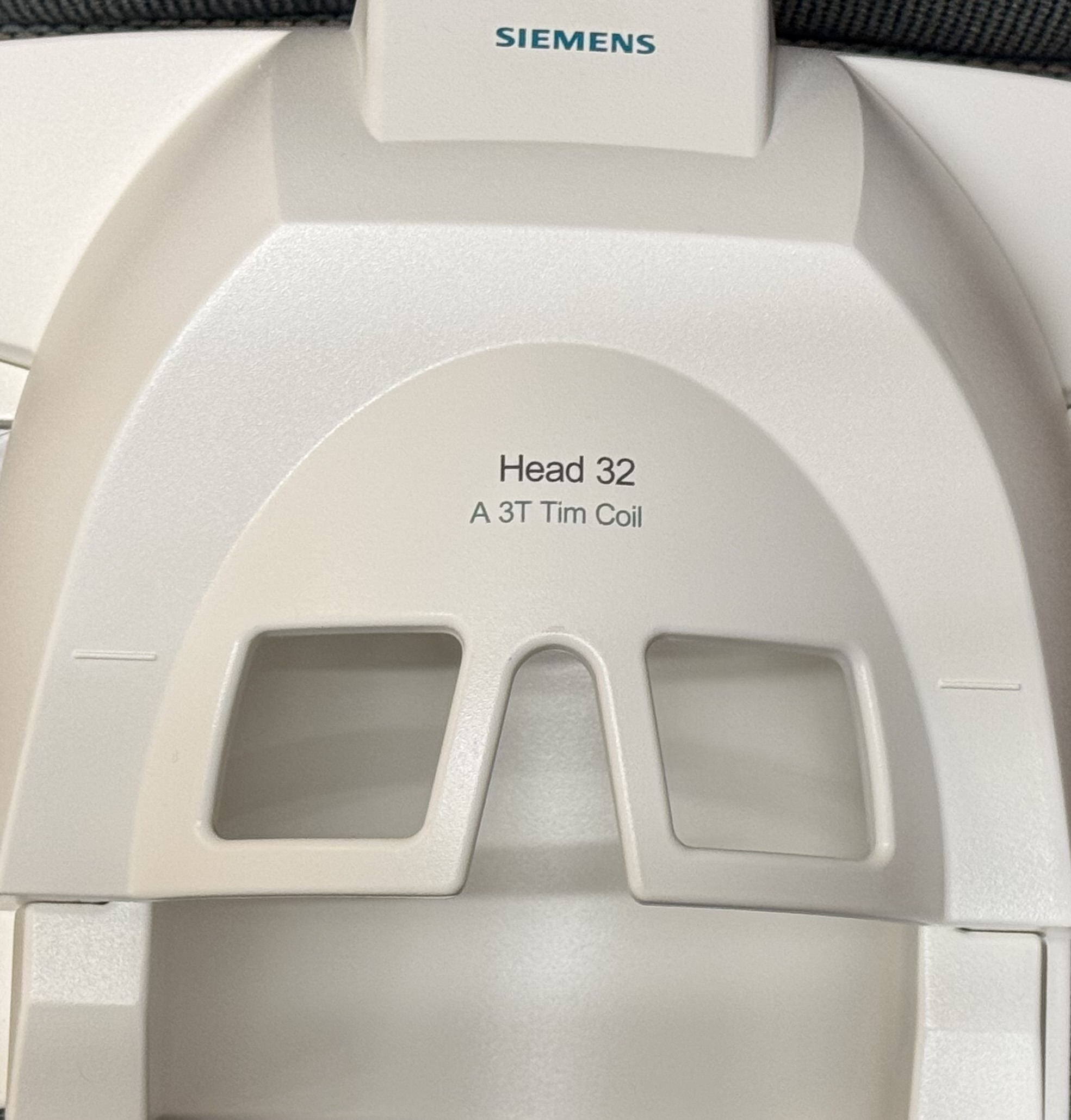
Figure 2: Frontal view of the Siemens 32-channel head coil as reference for precise mount adaptation.
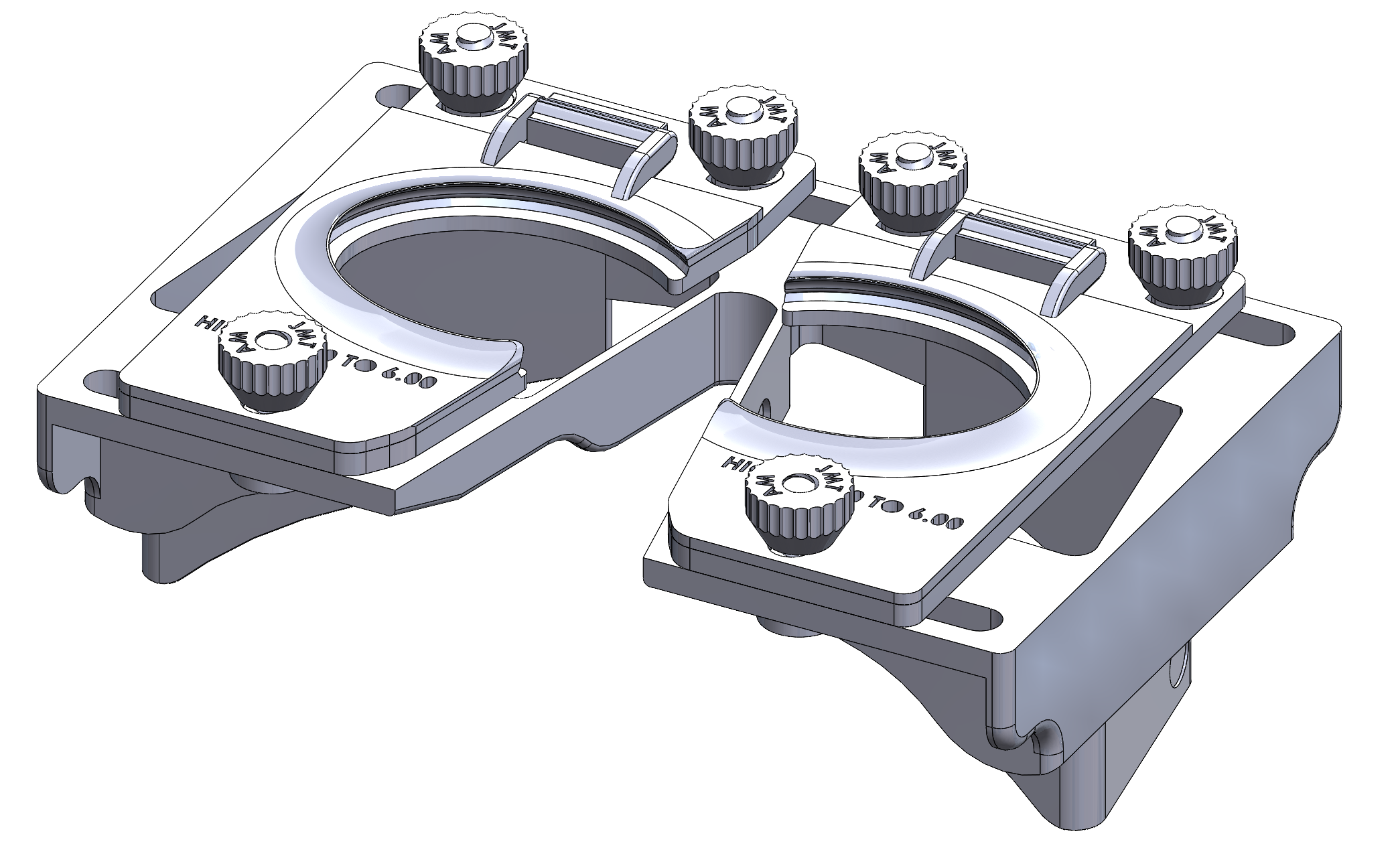
Figure 3: Trimetric rendering of the complete vision correction device, showing the interconnected configuration of mount, lens holders, and fasteners.
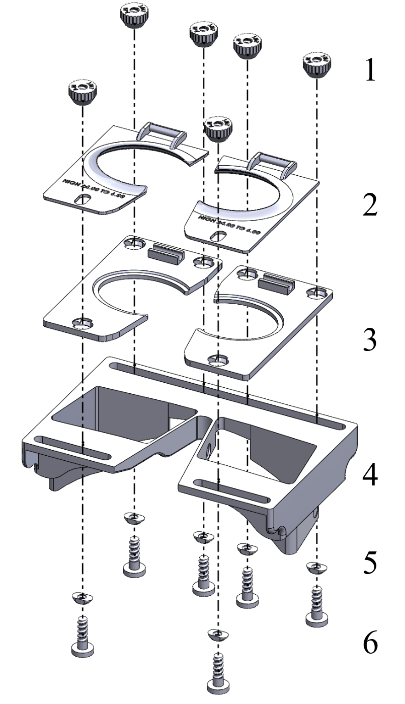
Figure 4: Exploded assembly illustrating all mechanical components required for practical device deployment.
Fabrication, Assembly, and Practical Considerations
The protocol supports rapid, low-cost manufacturing; the entire hardware set can be fabricated at a materials cost of approximately $25. Printing parameters are optimized for surface quality and robustness (0.08–0.20 mm layer height, high wall count, and 30% crosshatch infill for major structural elements). Assembly is straightforward: gel bumpers are affixed in specified recesses on the head coil mount to dampen acoustic vibrations, then lens holders and fasteners are combined, and finally the device mounts securely onto the coil.
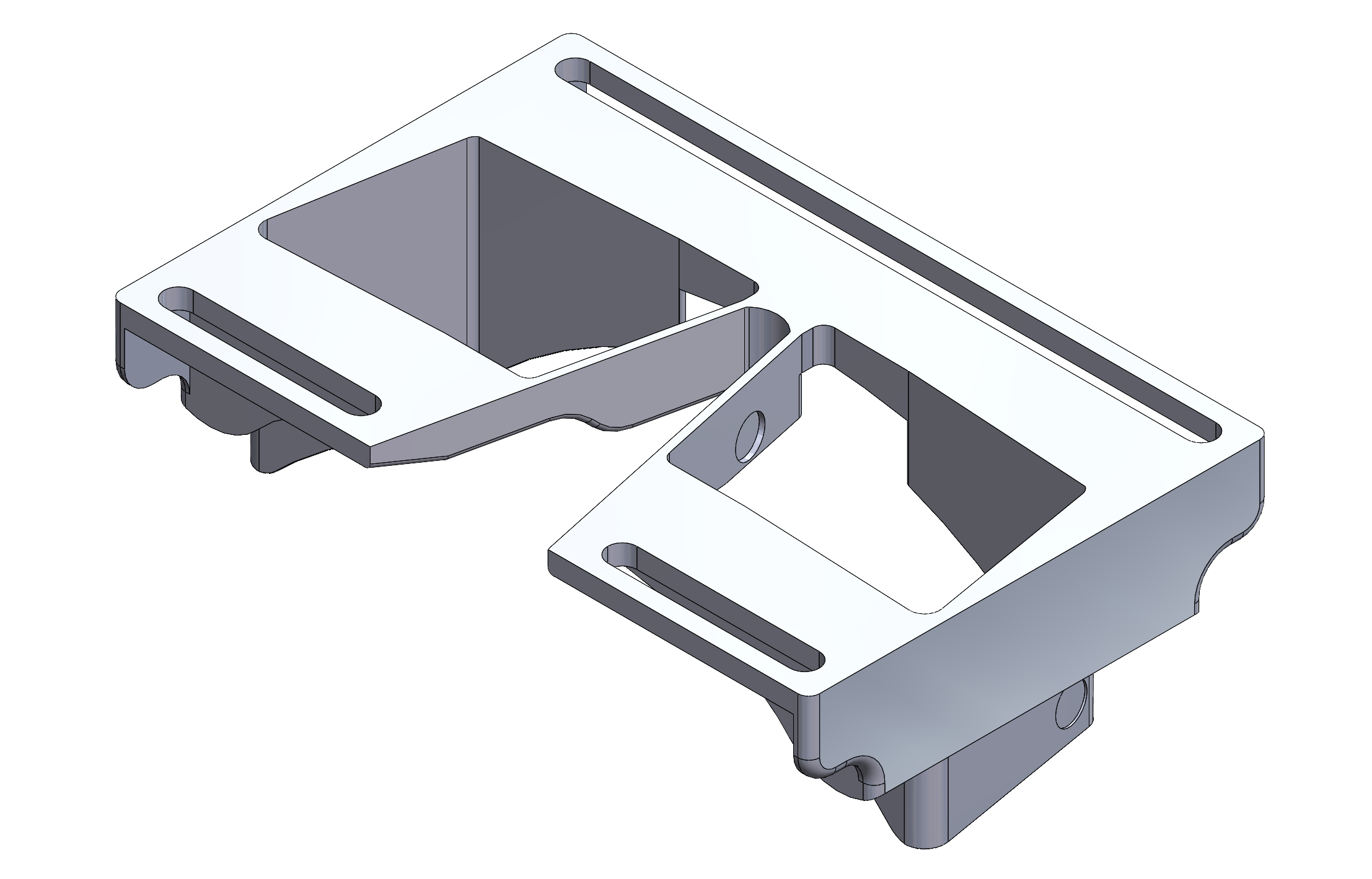
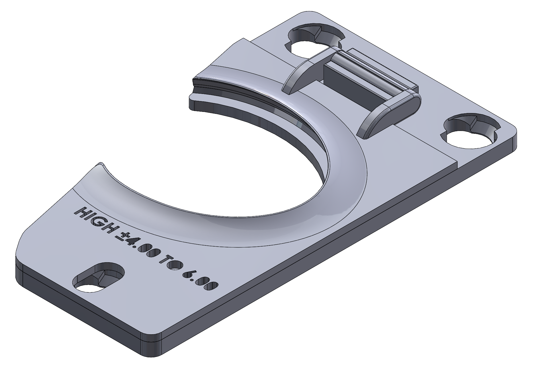
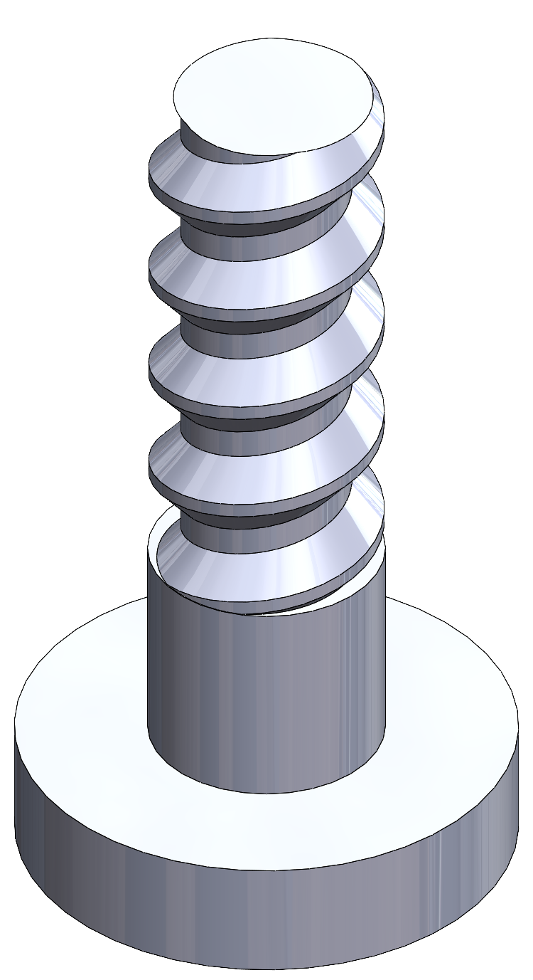
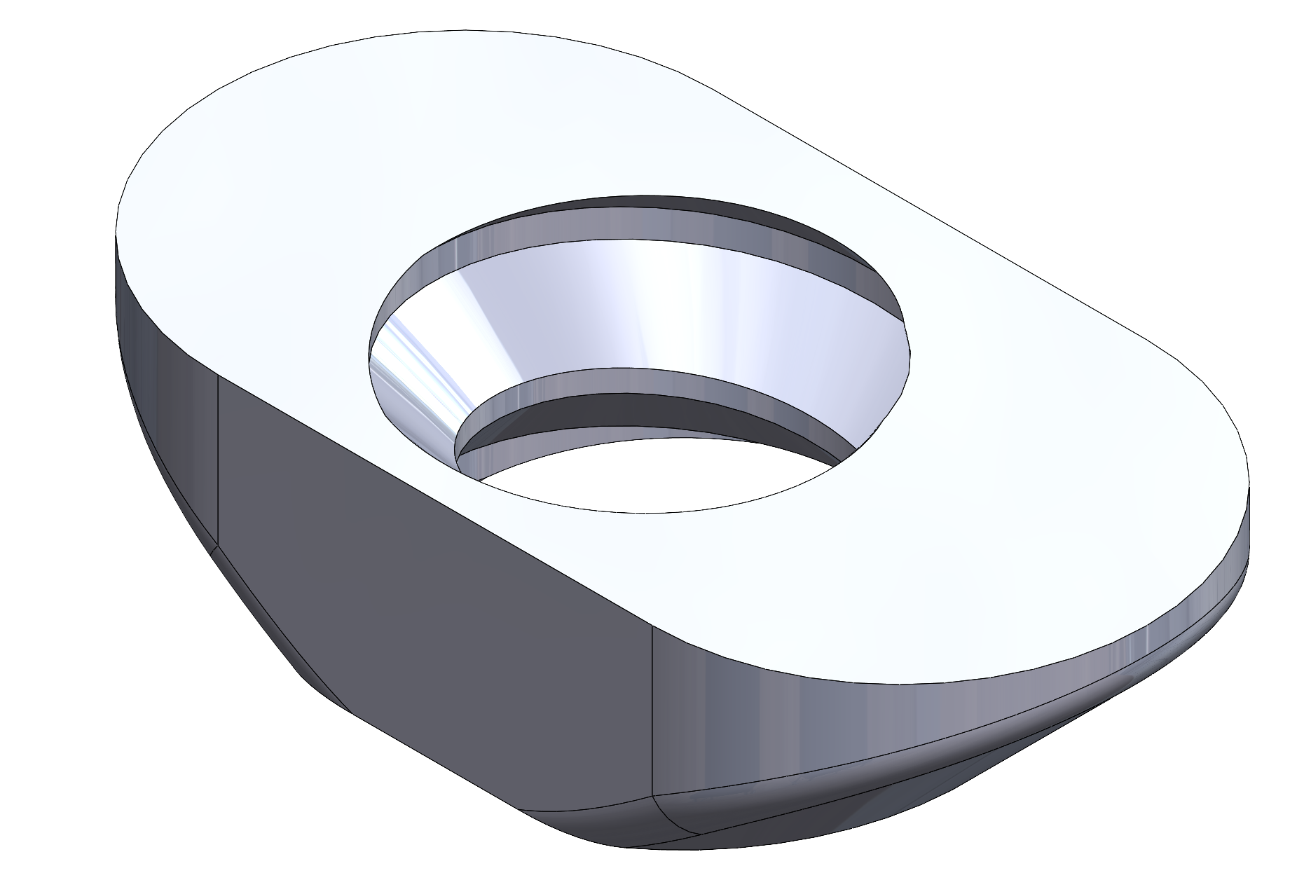
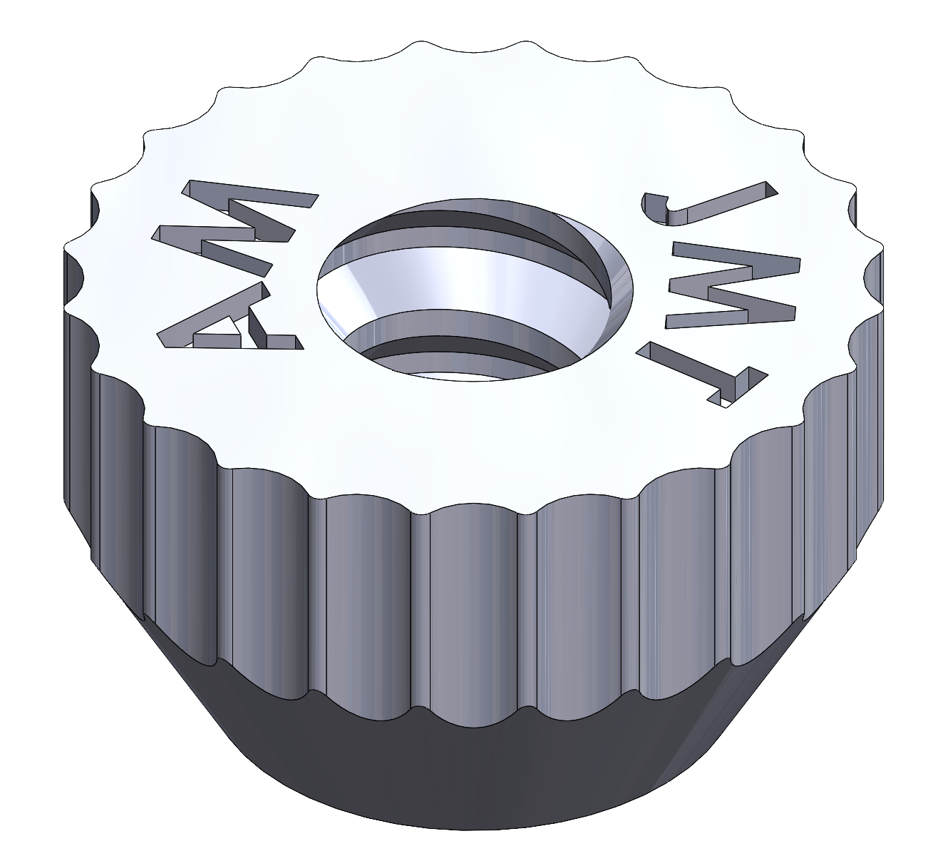
Figure 5: The isometric head coil mount with engineered recess locations for acoustic gel bumpers.
The design emphasizes ergonomic improvements: absence of head contact, ease of adjustment, and rapid lens exchange. Lens holders are secured by a hinge mechanism for quick insertion and removal. IPD adjustment is accomplished in situ, enhancing participant comfort and visual alignment accuracy.
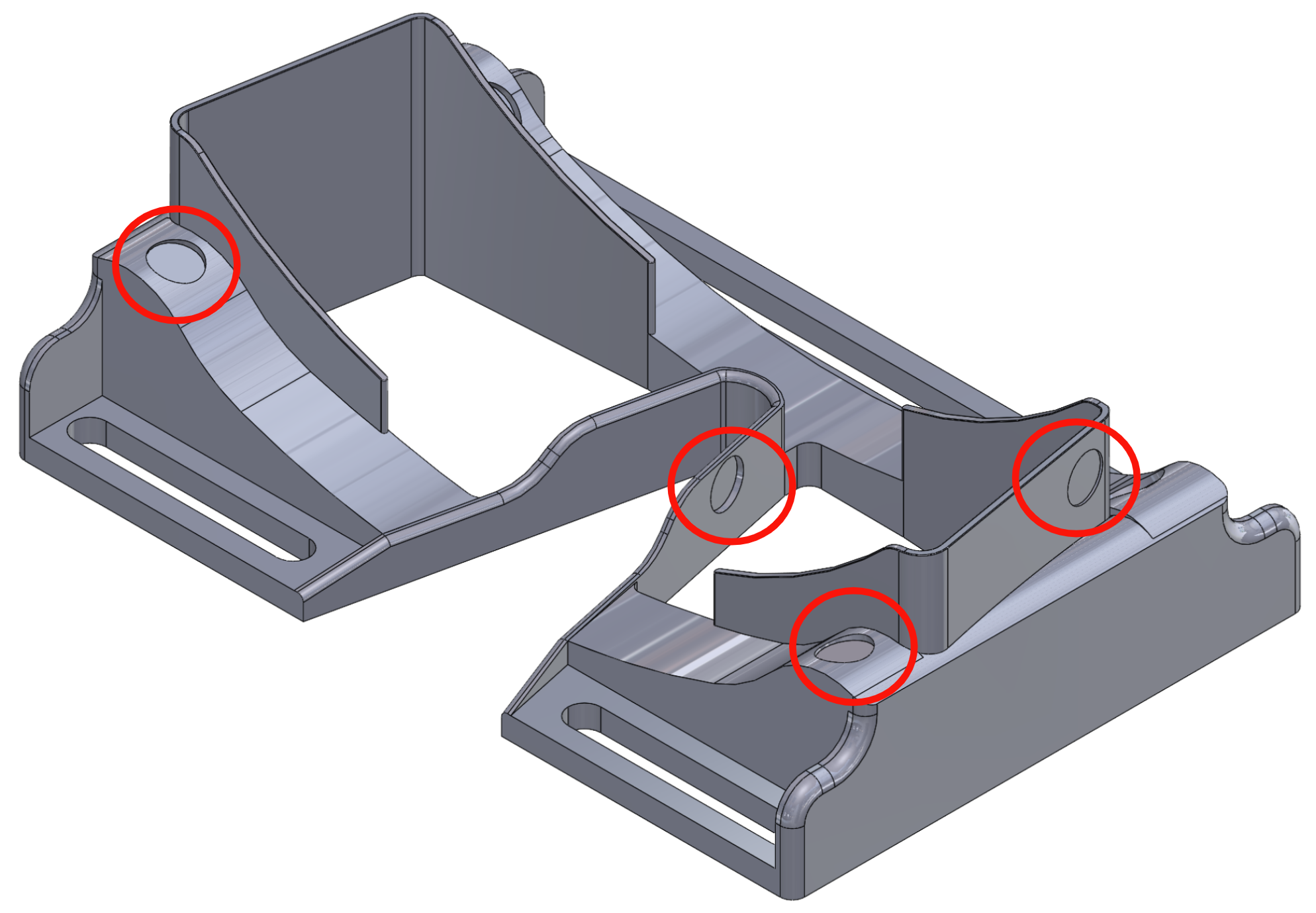
Figure 6: Bottom isometric view visualizing circular recesses for gel bumpers as vibration damping elements.
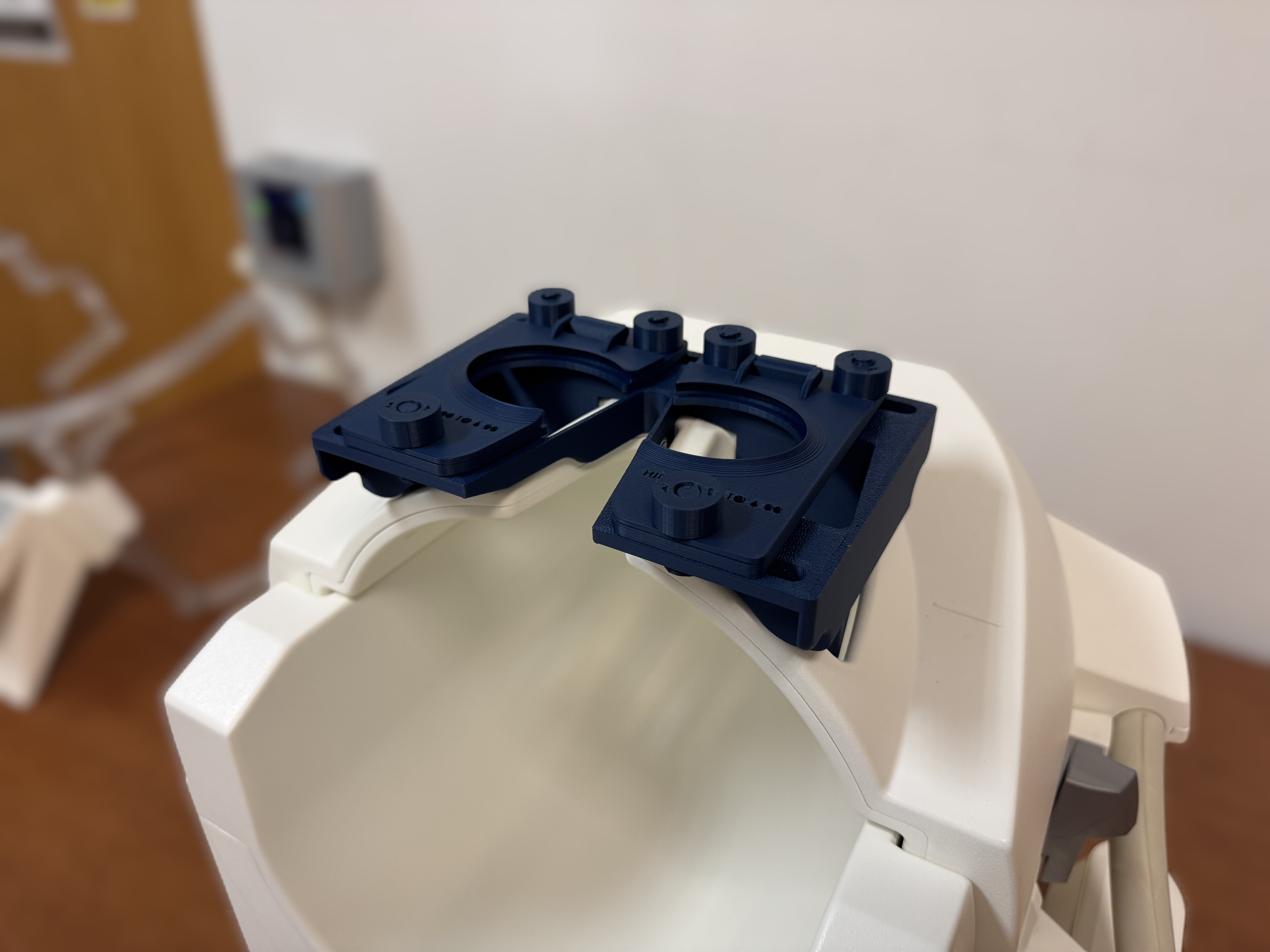
Figure 7: Device mounted on coil without lenses—a step in fit-checking prior to lens installation.
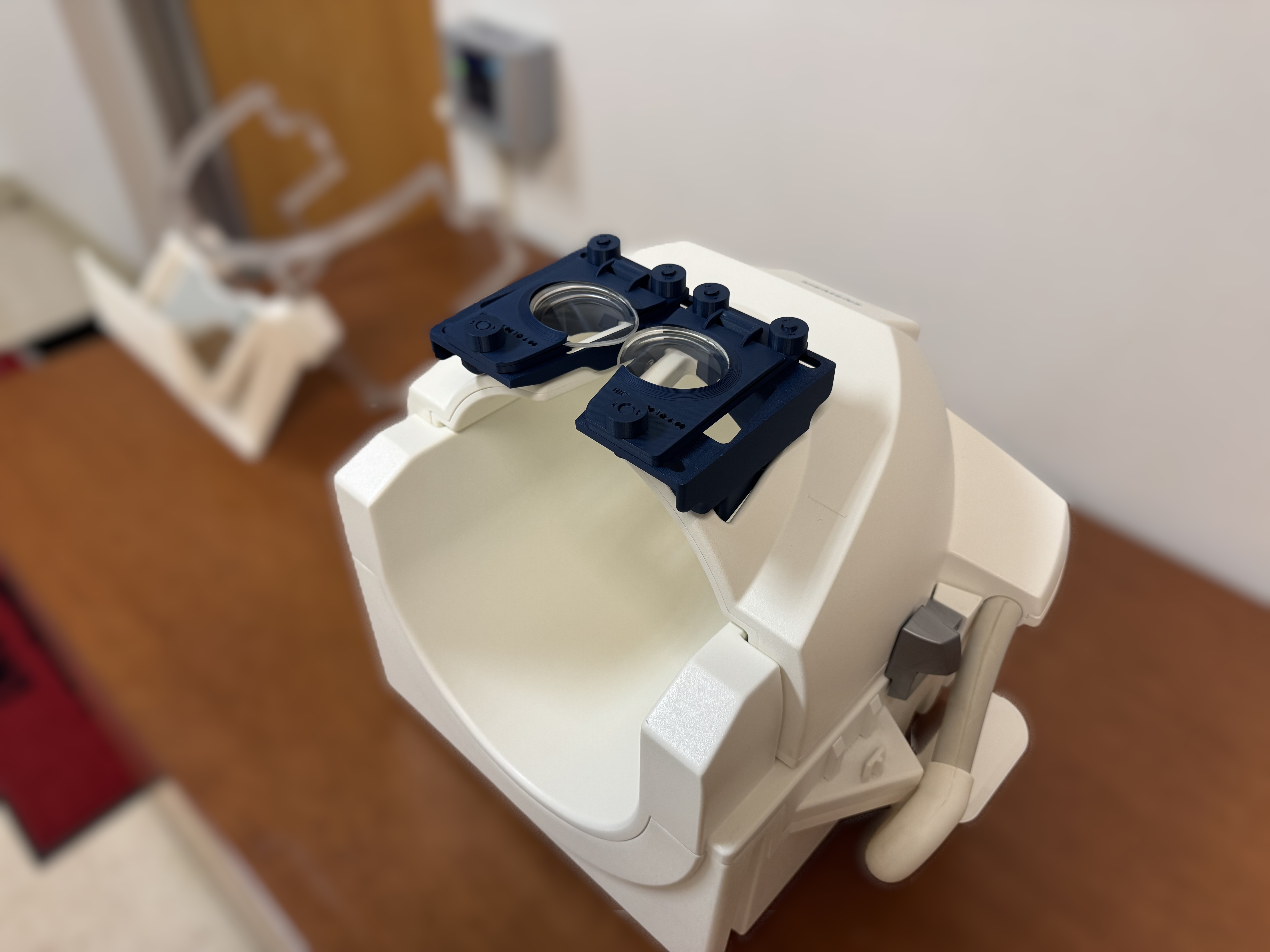
Figure 8: Device in operational configuration, with corrective lenses mounted and fixed in holders.
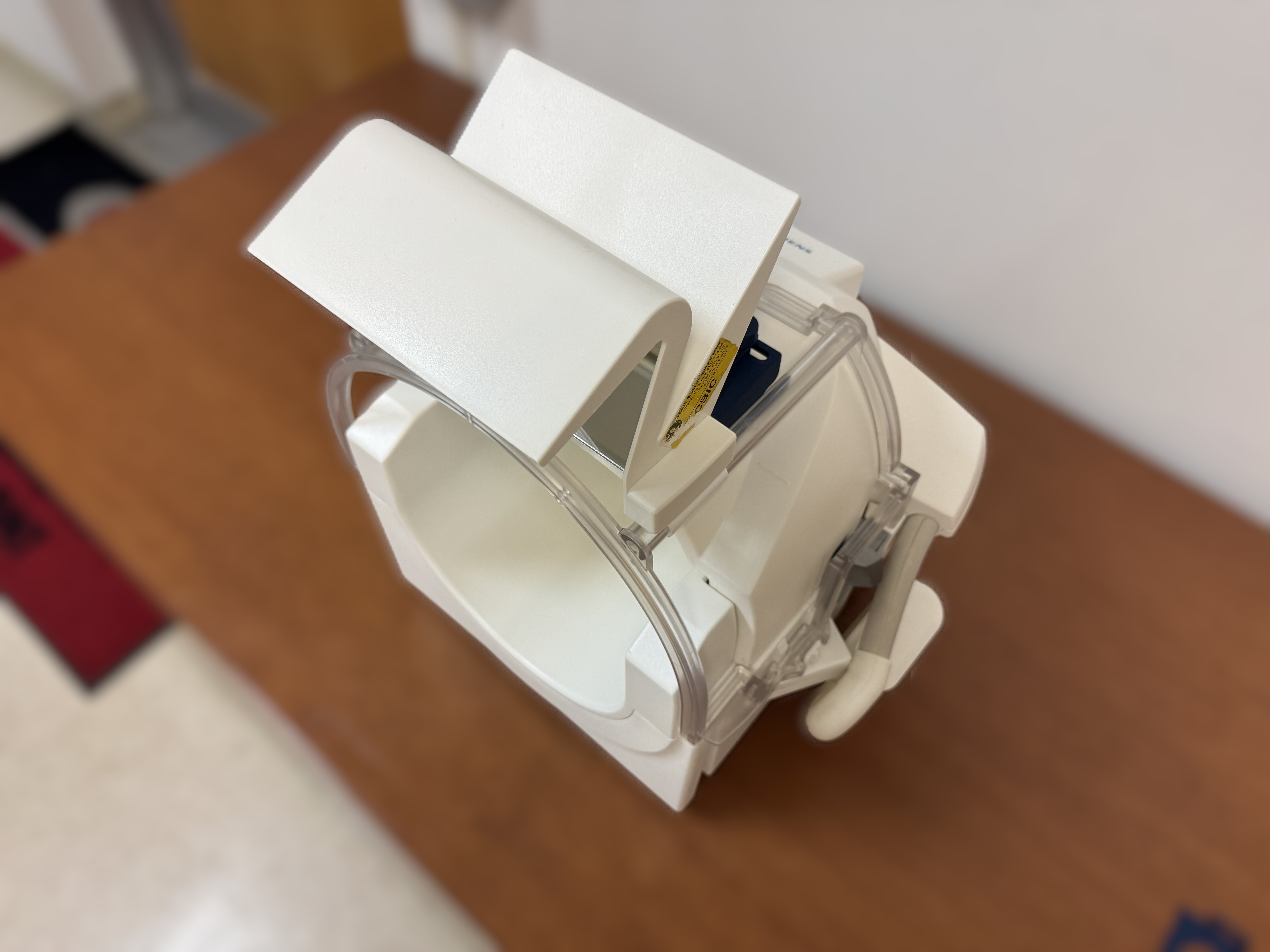
Figure 9: Full assembly (including mirror for visual stimulus reflection), ready for participant use in MRI scanner.
Modular Applications and Future Flexibility
The modularity of the platform facilitates rapid prototyping and adaptation to alternative lens set geometries, other head coil models (including General Electric and Philips), and additional experimental paradigms such as monocular occlusion. The entirely open-source CAD and print files (SLDPRT, STEP, STL) are provided under a CC BY-NC-SA 4.0 license, enabling community-driven optimization and adaptation.
Reflections, possible on some lenses under eye-tracking illumination, can be mitigated using anti-reflective coatings or by sourcing pre-coated lenses. The lens holders’ modularity further allows for tailored designs to specific research requirements.
Practical and Theoretical Implications
This device represents a technically rigorous, open hardware advance that directly addresses the persistent limitations of visual correction under MRI. The key technical contributions are: (1) fully adjustable IPD for improved visual fidelity, (2) rapid tool-less lens switching, (3) accommodation of ergonomic constraints for tight head coil geometries, and (4) full MRI compatibility—achieved through open-source, 3D-printable plastics and non-magnetic hardware.
On a practical level, the reduction in cost and simplification of fabrication protocols facilitate broader adoption in the MRI research community. On a theoretical level, systematic correction for refractive error variability—across a demographically representative subject population—enables higher data fidelity in vision science, neuroimaging, and psychoinformatics, and could reduce the confounds associated with compromised visual stimulus presentation.
Potential future directions include integration with eye-tracking systems (subject to further minimization of lens reflections), adaptation to specialized coils and higher-order optical corrections (e.g., for astigmatism or prism corrections), and extension to pediatric or clinical subpopulations with specialized ergonomic or refractive requirements.
Conclusion
This work delivers a fully functional, robust, and ergonomically optimized coil-mounted vision correction platform for MRI and fMRI research, significantly advancing flexibility and reproducibility in visual presentation protocols. The open-source dissemination model encourages iterative advancement by the neuroimaging instrumentation community, with direct positive impact anticipated for data quality and research subject comfort in vision-driven MRI experiments.

