- The paper introduces Tensor-ComBat, a Bayesian tensor response regression framework that incorporates spatial correlations to enhance scanner effect removal and preserve biological signals.
- It demonstrates significant improvements with higher correlations and lower RMSE compared to methods like ComBat, validated on ADNI-1 MRI scans.
- The approach enables principled uncertainty quantification and reproducible biological feature selection, setting a robust foundation for multi-site neuroimaging studies.
Introduction
Multi-site neuroimaging studies have become indispensable for increasing statistical power and reproducibility in neuroimaging research. However, these advantages are offset by pronounced scanner- and site-specific batch effects, confounding downstream analyses by introducing artifactual technical variability that can obscure or mimic biological signals. Existing harmonization approaches such as ComBat and adjusted-residuals estimators address batch effects by regressing out scanner influences, yet critically neglect spatial dependencies inherent in brain images. The paper "Spatially-informed Image Harmonization Results in Improved Scanner Effect Removal and Prediction" (2604.01546) introduces Tensor-ComBat (TC), a Bayesian tensor response regression framework that achieves harmonization by explicitly modeling spatial correlations within images.
Methodological Innovation: Bayesian Tensor-Based Harmonization
Tensor-ComBat (TC) generalizes the harmonization framework by incorporating spatial configuration via a low-rank PARAFAC tensor decomposition, enabling joint learning of both spatially-correlated mean (additive) and variance (multiplicative) scanner effects as well as biological covariate effects at the voxel-level. This fully Bayesian implementation facilitates principled uncertainty quantification, naturally supports high-dimensional parsimony, and enables the estimation of spatially distributed credible regions for scanner and biological effects—a property unattainable by empirical Bayes or ordinary least squares harmonization procedures.
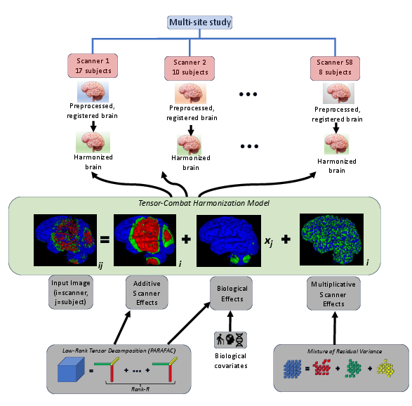
Figure 1: The pre-processing pipeline is illustrated in this Figure.
The model is applicable both cross-sectionally and longitudinally (with subject and temporal random effects), with harmonization feasible directly on volumetric images or on slice-wise decompositions to improve computational tractability and model expressivity. The harmonization step mirrors ComBat's adjustment but with parameters estimated from the Bayesian tensor model, realizing harmonized structural images with spatially-adaptive correction.
Experimental Paradigm: ADNI-1 MRI Harmonzation
TC harmonization was benchmarked on T1-weighted MRI scans from the ADNI-1 cohort (2108 images, 818 subjects, 58 scanners). Cortical thickness (CT) maps, a biomarker for AD-related neurodegeneration, were extracted after standardized registration and strict voxel selection pipelines, yielding 27,000-voxel downsampled brain volumes per image. Biological covariates including age, sex, clinical diagnosis, and APOE-4 status were incorporated in harmonization models; cognitive test scores (MMSE, RAVLT, ADAS) were reserved for downstream evaluation.
Quantitative Results
Enhanced Scanner Effect Removal
All harmonization methods improved pairwise similarity between scanner means compared to raw images, but TC-CS-Sl (cross-sectional, slice-wise) yielded significantly higher mean correlations (0.2% increase; p<2e−16) and lower pairwise RMSE (7% decrease; p=3.4×10⁻¹⁰) than ComBat. At the ROI level, improvements magnified to 0.8% increased correlation and 14.4% lower RMSE over ComBat. These gains are robust to adjustment for multiple comparisons.
Critically, TC's Bayesian implementation supports spatial inference: the highest scanner deviations localized to the cerebellum and bilateral inferior temporal/fusiform gyri, with up to 36% of scanner pairs displaying significant differences in cerebellum (Figure 2). This spatial specificity is unattainable with vectorized approaches.
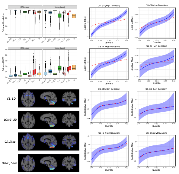
Figure 2: For distribution plots (left half), we define a region of ``high deviation'' as all voxels such that >10% of scanner pairs.
Residual analysis further supports scanner normalization efficacy: while both TC and ComBat reduced scanner-effect ANOVA rejections to <0.5% of voxels, TC delivered superior fit (R2=0.75 vs. p<2e−160 for ComBat, p<2e−161) and substantially fewer residual outliers.
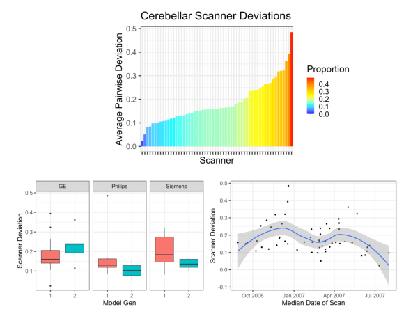
Figure 3: Generation 1 Models: Genesis Signa, Signa Excite (GE), Gyroscan NT, Gyroscan Intera, Intera (Philips), Sonata, SonataVision, Symphony (Siemens). Generation 2 Models: Signa HDx (GE), Intera Achieva, Achieva (Philips), SymphonyTim, Avanto, NUMARIS/4 (Siemens)
Scanner deviations were modulated by hardware generation (notably for Siemens, significant reduction from generation 1 to 2), with deviation magnitude correlating with scanner sample size and timing.
Superior Biological Signal Prediction
For biological covariates included in harmonization, TC-CS-Sl harmonized images produced the lowest cross-validated out-of-sample errors. Age prediction RMSE was lower by a significant margin across all comparator algorithms (p<2e−162 vs. raw data, p<2e−163 vs. ComBat). Similar significant gains were observed for diagnoses and APOE-4 status, consistently confirmed across full and 50% subsampled training sets.
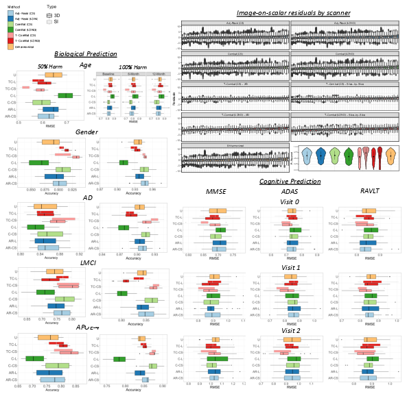
Figure 4: Biological (left) and cognitive scores (bottom right) prediction using unharmonized and harmonized images, fit to age, gender, cognitive syndrome diagnosis, APOE4 and cognitive scores. And image-on-scalar residuals by scanner.
For cognitive variables (excluded from the harmonization model), predictive gains were present but attenuated: RMSE for MMSE improved for baseline images (p<2e−164), while ADAS prediction was significantly improved at all timepoints, particularly at 1-year follow-up (RMSE reduction of p<2e−165 over ComBat). Longitudinal harmonization variants showed diminished gains, likely due to over-parameterization at limited timepoints.
Reproducibility of Biological Feature Selection
The dice coefficient between significance maps (on biological covariate regression) was markedly higher for TC (mean Dice 0.96 for age, 0.79 for LMCI) compared to ComBat and other methods, particularly for AD-relevant effects (Figure 5). Reproducibility was robust across split-halves and replicate analyses.
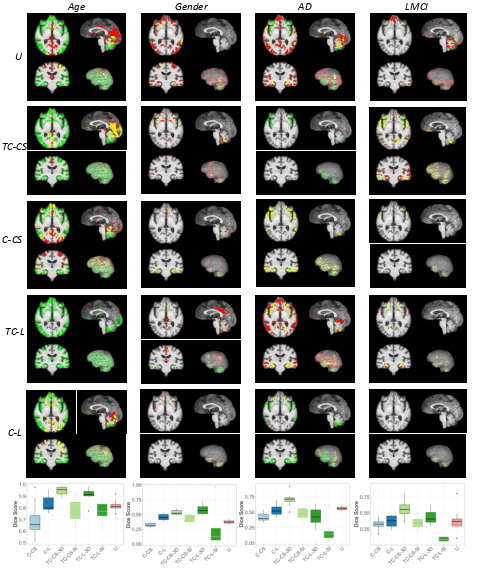
Figure 5: The overlapping significance maps and Dice scores for age, gender, cognitive syndrome diagnosis (AD or MCI).
Statistical Significance
All primary result claims were supported by rigorous statistical testing (t-tests and multiplicity corrections with Benjamini–Hochberg), buttressed by comprehensive p-value tables (see Supplementary Materials).
Theoretical and Practical Implications
TC demonstrates that explicitly modeling spatial dependencies drives consistent gains in harmonization. By resolving scanner effects more effectively—with detailed quantification of uncertainty—while amplifying relevant biological signals and ensuring high reproducibility, TC surpasses vector-based harmonization approaches on all consequential metrics in large, multi-site neuroimaging studies.
The Bayesian tensor methodology generalizes to other imaging modalities and multi-dimensional biomedical arrays, supports scalable high-dimensional inference, and enables uncertainty quantification on spatially distributed effects—capabilities directly applicable to GWAS imaging phenotypes, longitudinal trials, and large-scale population studies.
Future Directions
The high model flexibility comes with increased computational cost, particularly for longitudinal deployment given current MCMC algorithms and the requirement for more per-subject timepoints to avoid overfitting. Future research into scalable variational approximations, adaptive rank selection, and automatic spatial hyperparameter inference (e.g., via hierarchical spatial process priors) could further improve TC scalability and generalizability. Empirical extensions to non-Gaussian neuroimaging endpoints, multi-modal image fusion, and integration with federated or privacy-preserving harmonization settings are also natural directions.
Conclusion
Bayesian spatially-aware harmonization, as instantiated in Tensor-ComBat, achieves superior scanner effect removal, biological signal preservation, and effect reproducibility compared to leading methods such as ComBat. This framework enables robust cross-scanner inference and downstream analysis for large-scale multi-site neuroimaging datasets, and sets a strong methodological foundation for harmonization in high-dimensional spatially-correlated biomedical data.