- The paper demonstrates that macroscopic fiber architecture predominantly governs active heart mechanics, while micro-disarray mainly affects contraction efficiency.
- It employs advanced imaging and Helmholtz filtering to decouple global organization from local fiber disarray in murine heart models.
- The study reveals that surrogate rule‐based models inadequately capture local strain patterns compared to detailed, experimentally calibrated fiber architectures.
The Functional Impact of Myofiber Macroscopic Organization and Disarray in Murine Heart Computational Models
Introduction
This paper investigates the respective roles of macroscopic myofiber architecture and microscopic fiber disarray in computational models of murine cardiac electromechanics, employing a biventricular model calibrated with high-resolution mesoscopic optical imaging. The study addresses the limitations of surrogate rule-based methods (RBMs) and examines effective modeling strategies for fiber disarray, with a focus on functional and mechanical consequences across electrophysiology, passive mechanics, and active contraction.
Methods: Measurement and Decomposition of Fiber Architecture
The myofiber field was measured using optical tissue clearing and high-resolution mesoSPIM microscopy, permitting quantification of both the macroscopic helical organization of cardiomyocyte bundles and local orientation variability at a resolution of $\SI{96}{\mu m}$. The paper formalizes a reference frame transformation relating fiber angles (α,γ,β) to the myocardial coordinate system and implements a Helmholtz low-pass filter to decouple macroscopic architecture (low frequencies) from disarray (high frequencies).
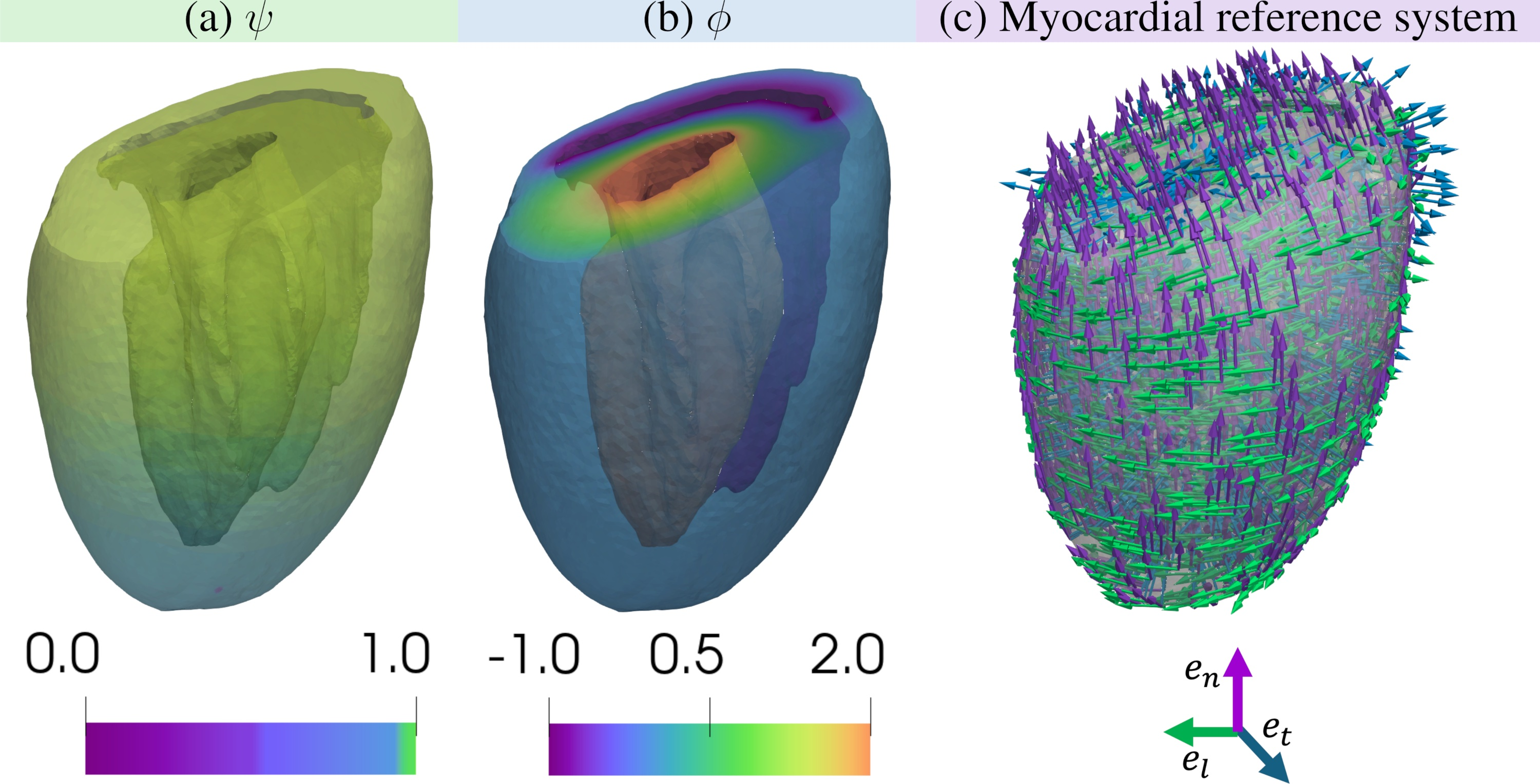
Figure 1: Visual schema illustrating the transformation from myocardial to fiber reference; key for parametrizing fiber orientation.
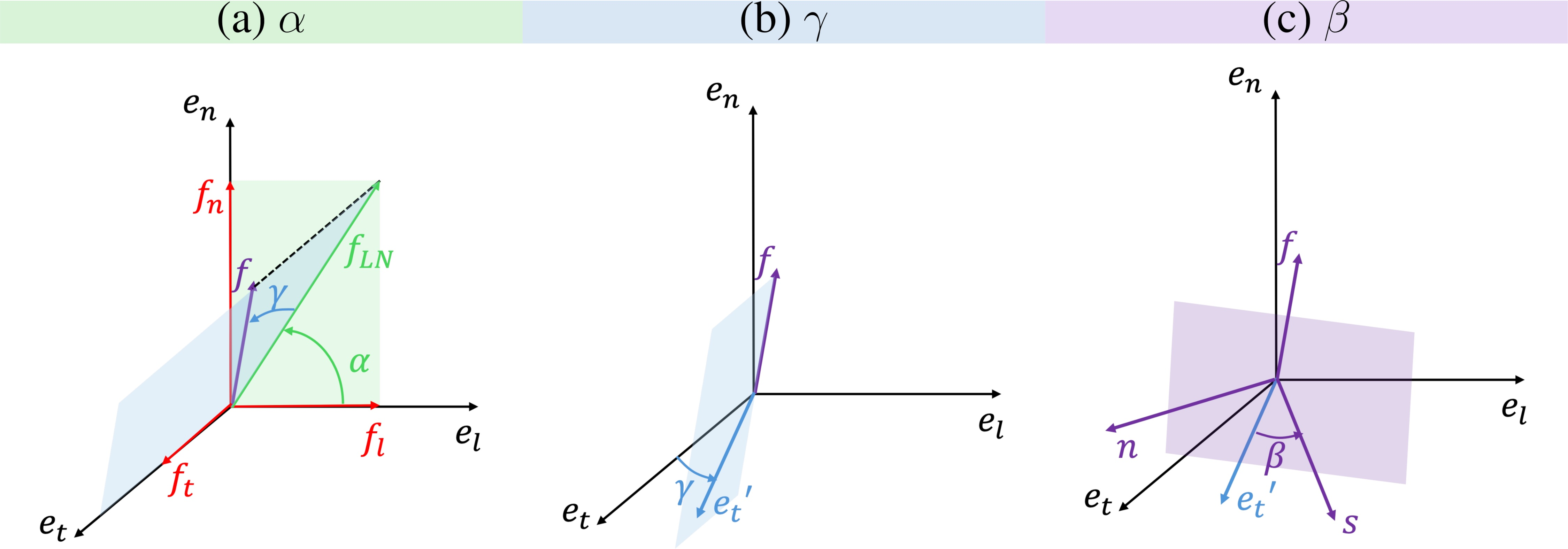
Figure 2: Construction and visualization of the myocardial reference system, which forms the backbone for mapping fiber orientation with respect to ventricular geometry.
Comparisons are made between the measured field, varying degrees of regularization (i.e., filter length ℓ), and a Laplace-Dirichlet RBM (LDRBM) generated field.
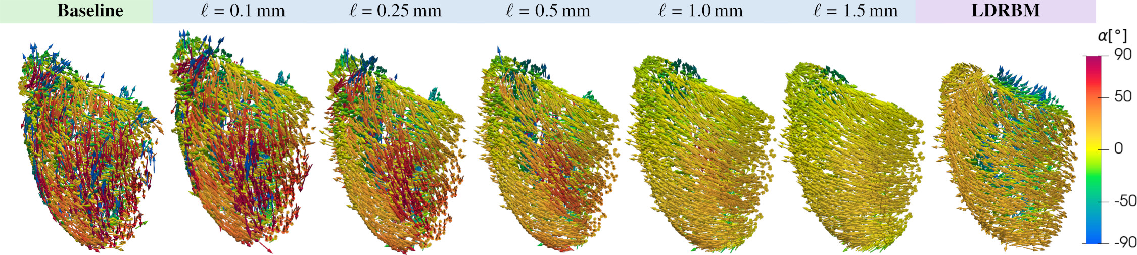
Figure 3: Fiber field visualization under increasing regularization; extreme smoothing or RBM-based surrogation highlights the loss of microscale features.
Statistical analysis of the angular distribution (Figure 4, Figure 5, Figure 6) demonstrates marked transmural heterogeneity in α but relatively small variance in γ, with smoothing reducing overall angular dispersion.
Electromechanical Model and Calibration
The multi-physics model comprises:
- Electrophysiology: An eikonal-diffusion approach, using transversely anisotropic conductivity tensors aligned with fiber orientation.
- Mechanics: Hyperelastic constitutive passive law (Usyk potential) and a detailed active stress (RDQ20-MF model), with contractile strength modulated by the crossbridge stiffness parameter aXB.
- Circulation: 0D cardiovascular system models coupled with the mechanics via proper boundary conditions and forward Euler integration.
Calibration proceeds by iterative adjustment of model components using specimen-specific data: activation maps, calcium/force traces, and reference pressure-volume loops extracted from both experiments and the literature.
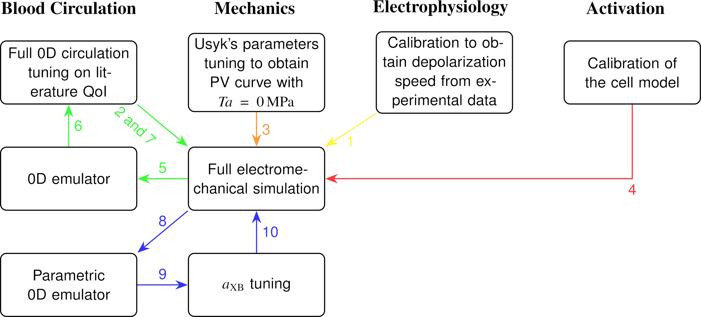
Figure 7: Workflow for progressive, multi-component calibration of the closed-loop biventricular electromechanical system.
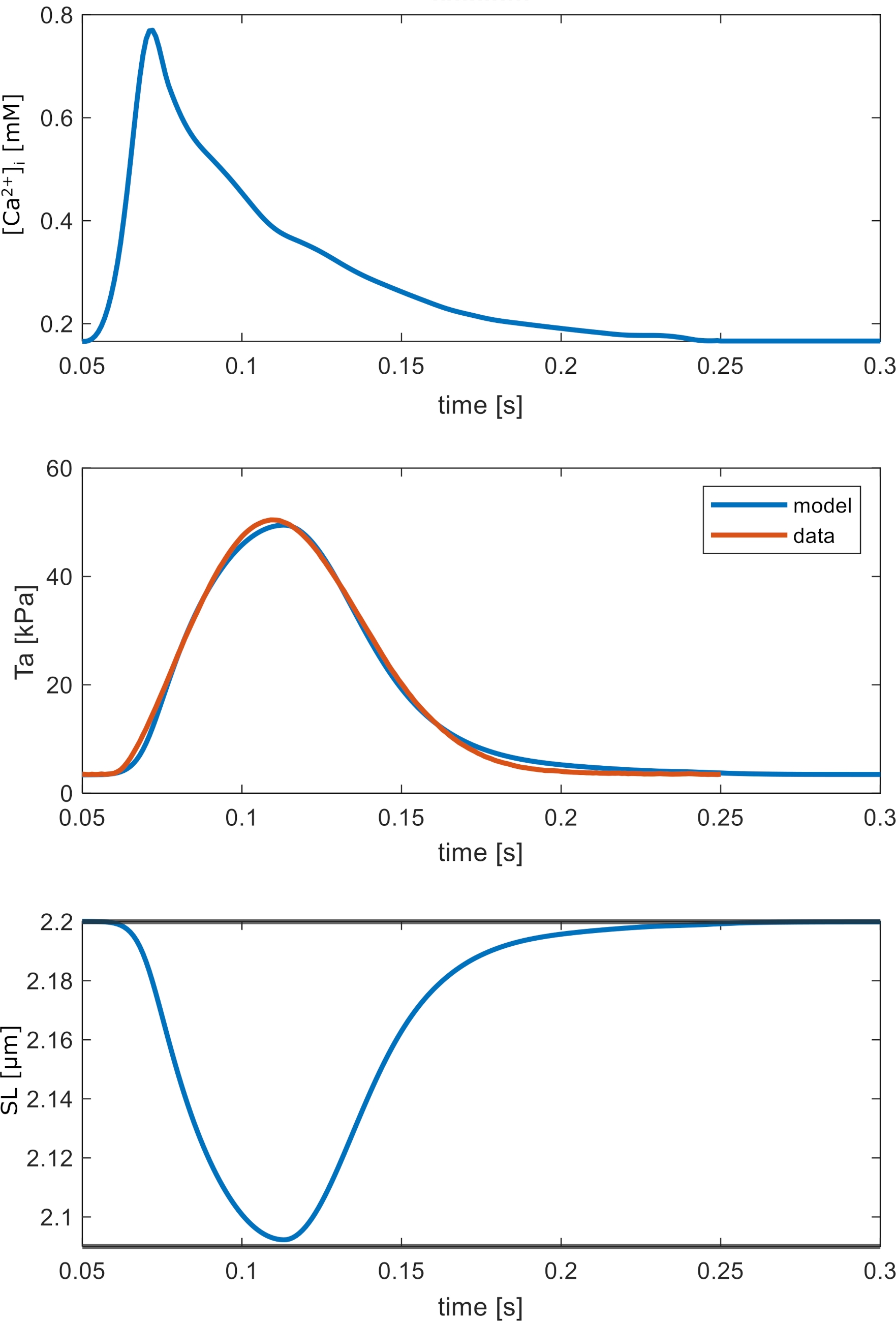
Figure 8: Calibration of the active force generation model; model reproduces key characteristics of experimental twitch force and calcium transients.
Results
Electrophysiology
The propagation of the activation wavefront is only marginally influenced by fiber disarray. Disarray increases isotropy in conduction, while both regularized and RBM fields yield more anisotropic, fiber-aligned propagation but produce similar global activation patterns and timings.
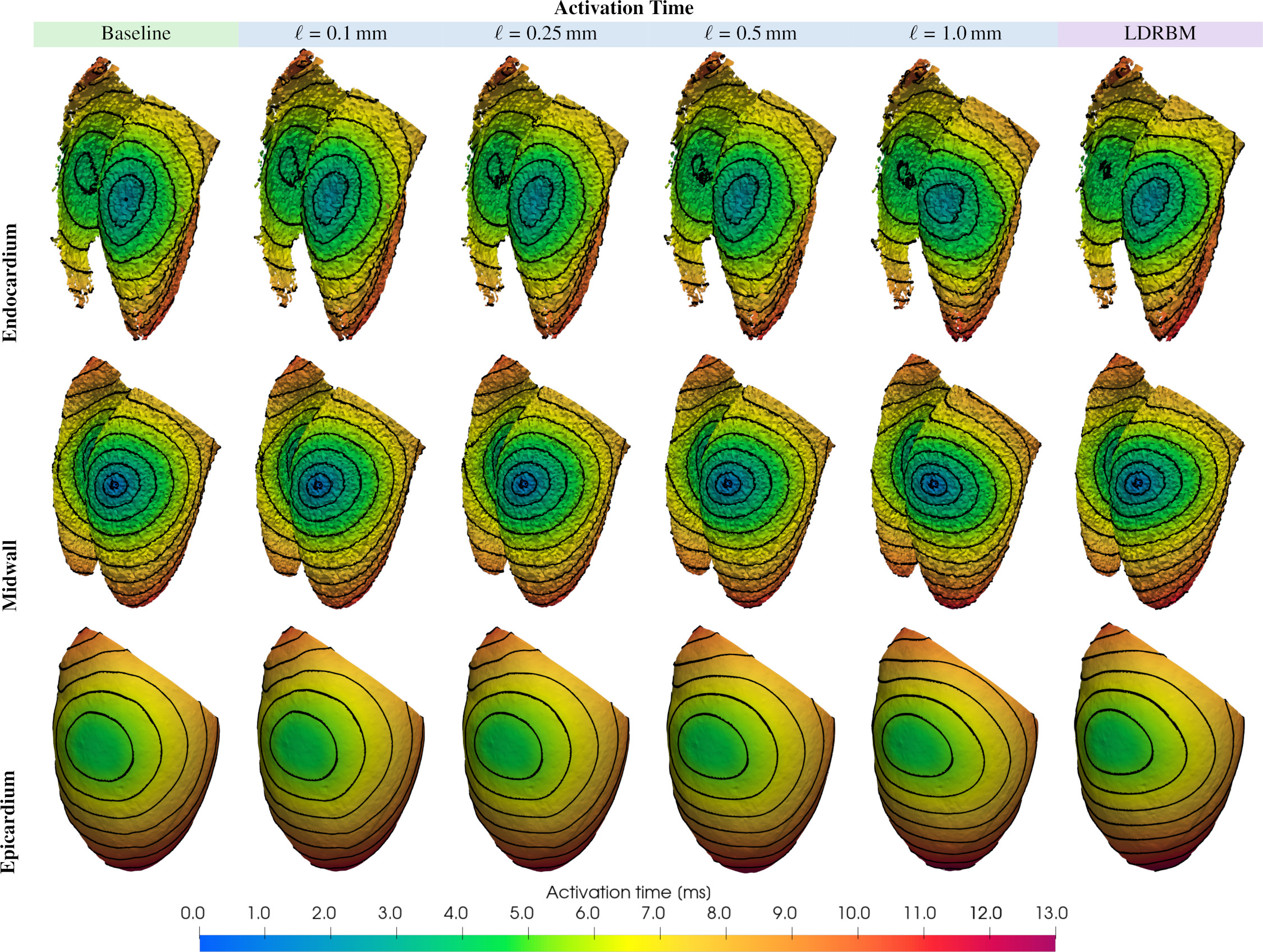
Figure 9: Isochronal maps indicate the influence of the fiber field on 3D conduction; regularization sharpens the structural anisotropy in wavefront directionality.
Passive Mechanics
Macroscopic architecture drives chamber compliance: the difference in ventricular volume at standardized pressure comparing experimental and LDRBM fields is approximately 4%. By contrast, passive mechanical response is almost insensitive to micro-disarray: smoothing from ℓ=0 to $\SI{0.25}{\text{mm}}$ changes volume by ~0.4%.
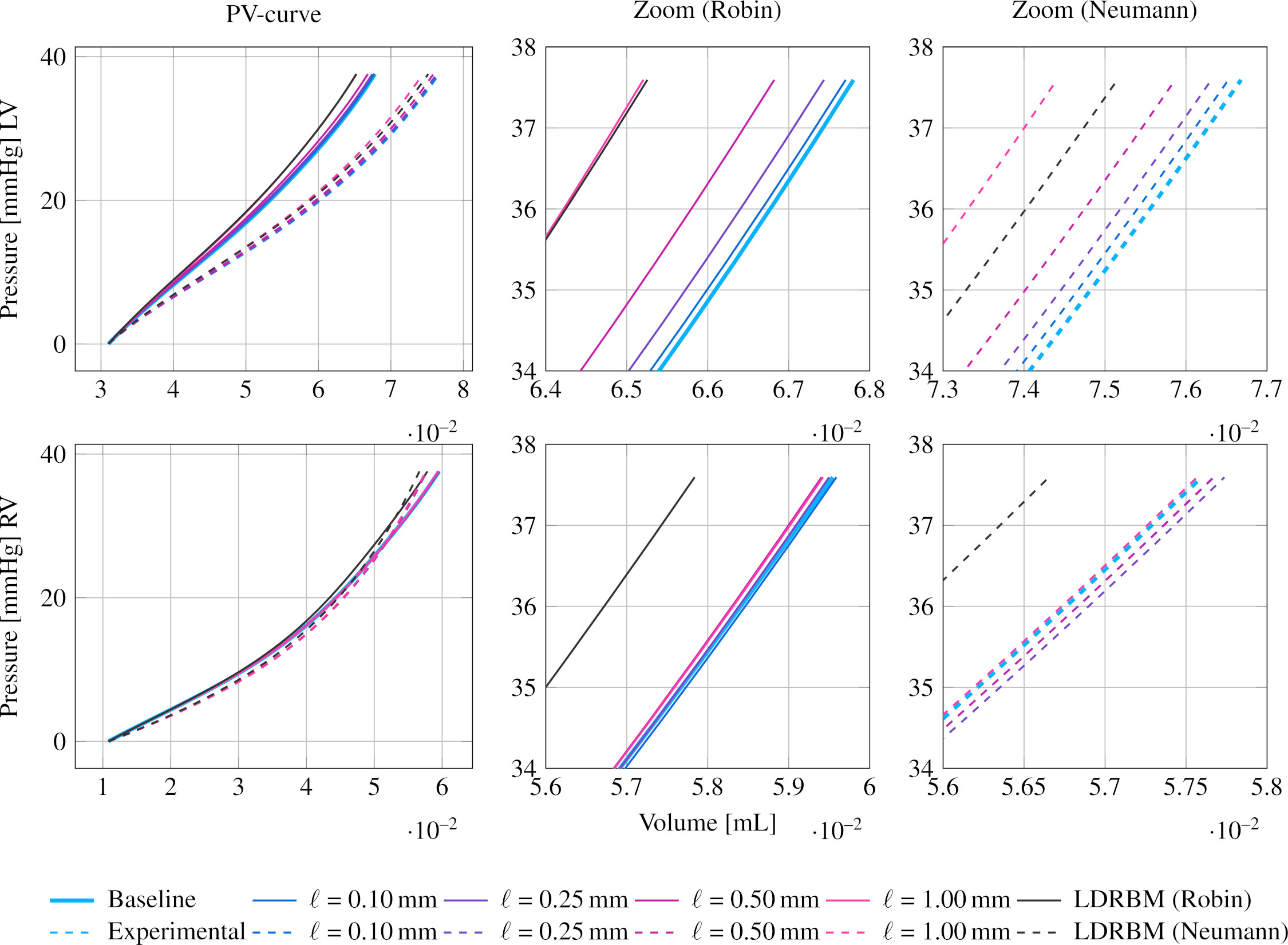
Figure 10: PV curves under passive inflation for LV and RV under different fiber architectures and boundary conditions.
Active Mechanics and Electromechanical Function
Active contraction is highly sensitive to both macroscopic architecture and fiber disarray. Moderate smoothing (removing only short-range disarray, preserving long-range organization) maximizes ejection fraction (EF); excessive smoothing (masking helical structure) or LDRBM fields either produced unphysiologically strong contraction (EF>90%, mesh failure) or inefficient contraction, confirming that both loss of order (too much disarray) and loss of helical structure are deleterious.
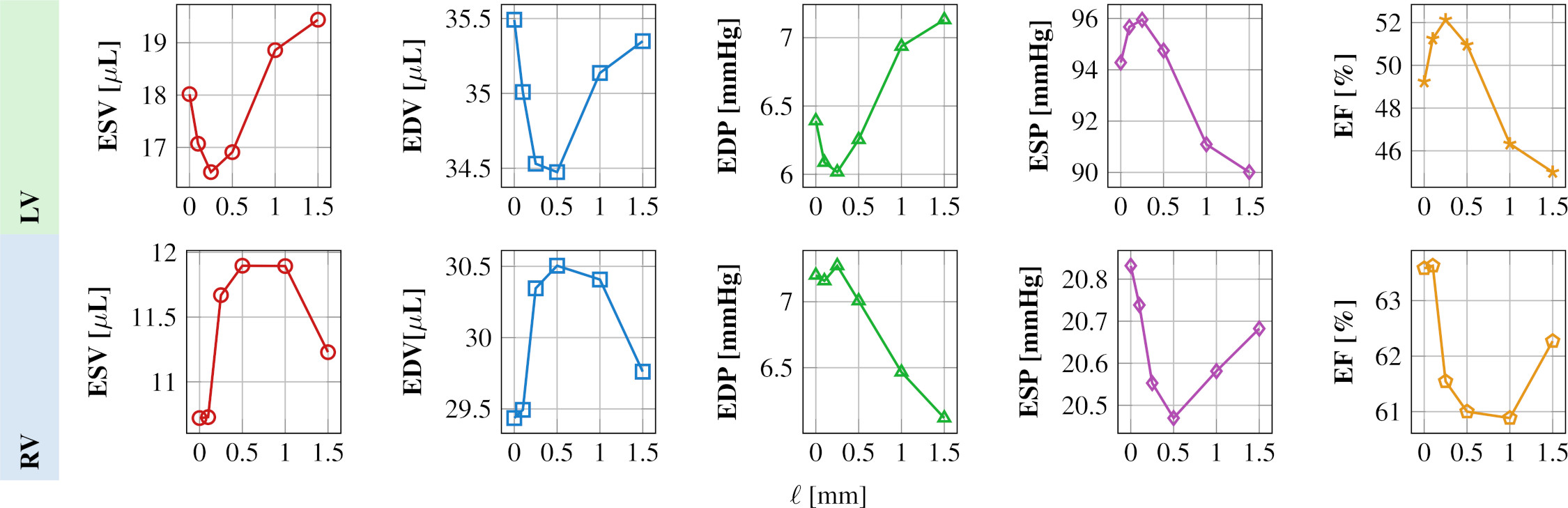
Figure 11: Hemodynamic QOIs (EDV, ESV, EF, etc.) as a function of fiber regularization radius, highlighting a non-monotonic relationship.
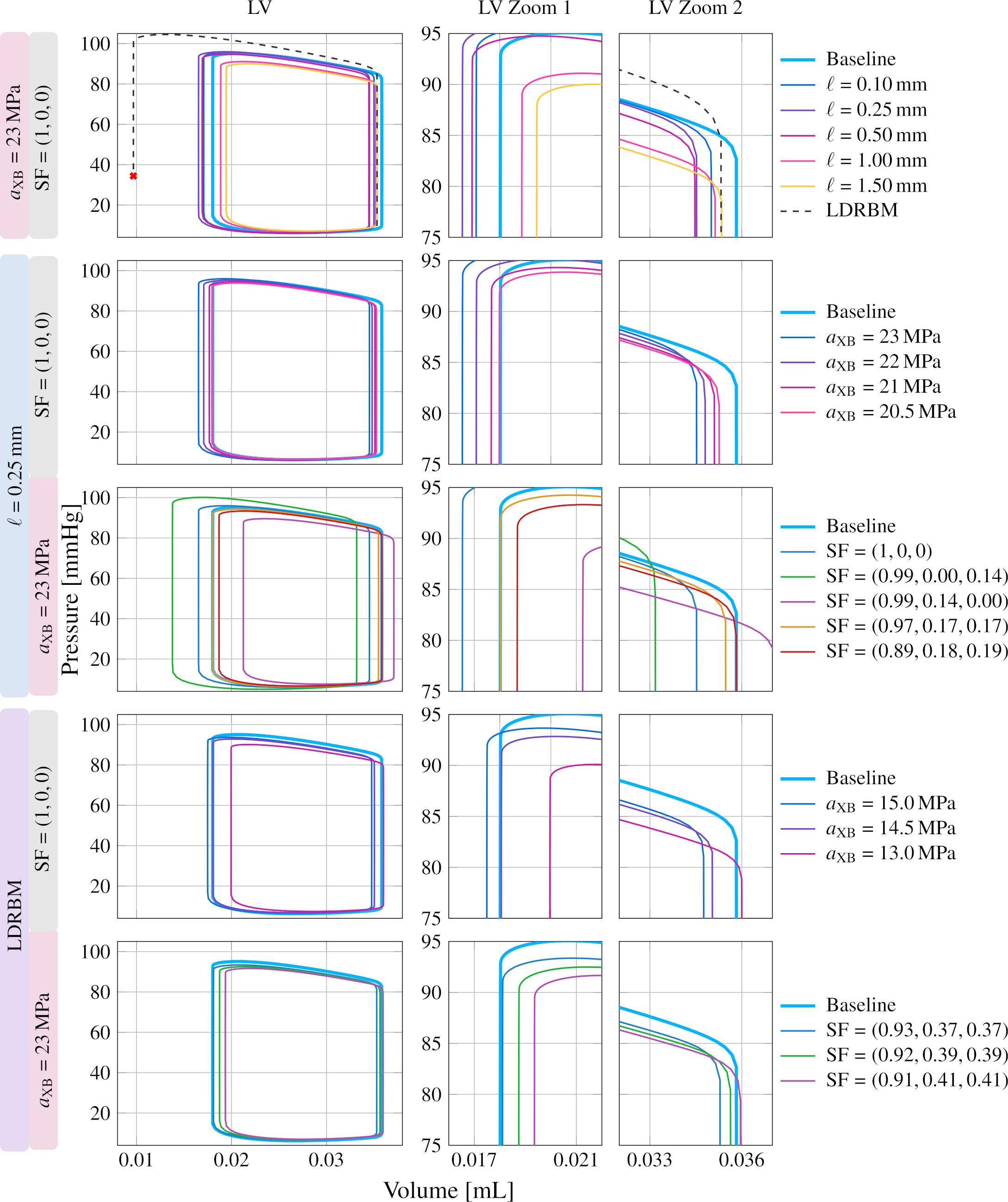
Figure 12: PV loop superposition for LV with experimental, regularized, and LDRBM-based fields. Only moderate regularization reproduces physiological contractility.
Two modeling strategies were assessed for capturing the effects of disarray in the absence of high-resolution data:
- Contractility reduction (aXB): Lowering peak active tension matches EF and PV loops effectively and recovers both global and local (strain) measures with the regularized architecture.
- Cross-fiber activation (nonzero sheet/cross Stress Factors): Enables matching the PV loop envelope, but fails to recover local strain heterogeneities and introduces non-physiological distributions.
Explicitly, for the best-fit case, a 13% reduction in (α,γ,β)0 reproduces the experimental PV loop with a regularized field. When RBM fibers are used, even stronger reduction is necessary, but the match is less faithful.
Strain Analysis
The first three invariants of the Green-Lagrange strain expose the limitations of surrogate modeling. Only moderate contractility reduction with regularized fibers restores local patterns of hydraulic and distortional strain; artificially aligning activation along cross-fiber directions fails to replicate experimental spatial strain distribution.
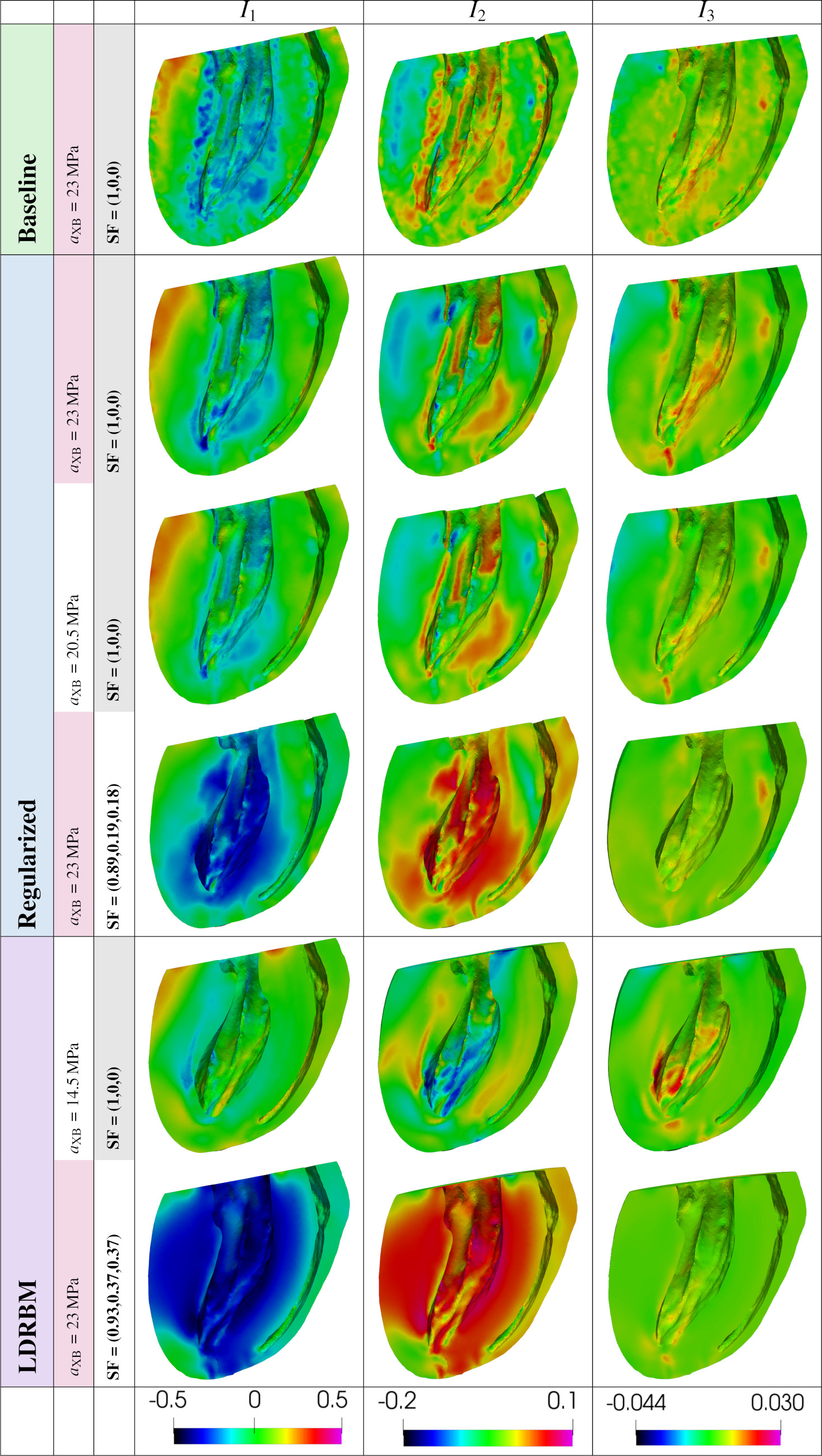
Figure 13: Analysis of tensor invariants confirms that only proper macroscopic organization, not surrogate models, can recapitulate local deformation features.
Discussion
The major conclusions are:
- Macroscopic architecture dominates active mechanics and chamber function; micro-disarray (within physiological range) has minor contributions except for contraction efficiency.
- Passive and electrophysiological behaviors are insensitive to fiber disarray, underscoring the adequacy of mean field approaches for these modules under physiological conditions.
- Conventional surrogate models are fundamentally limited: while global indices (e.g., EF, PV loops) can be mimicked either by tuning contractility or introducing cross-fiber activation, only a direct representation of macroscale organization allows correct spatial reproduction of strain and deformation.
- The methodology enables, for the first time, systematic decoupling of architectural scales in experimental data and direct quantitative assessment of their electromechanical impact.
- Excessive regularization or neglect of helical architecture (as in many RBMs) yields unphysiological global function (e.g., mesh instability, extreme EF).
- These results underline the need for improved upscaling techniques connecting explicit microstructure with effective active stress models, especially for applications targeting tissue-level phenomena (e.g., strain patterns, arrhythmogenic substrate mapping).
- Applicability to the atria, to pathological substrate (fibrosis, infarct, etc.), and inter-species extrapolation remain to be systematically explored.
Conclusion
This work rigorously demonstrates that in biventricular murine heart models, accurate reproduction of macroscopic myofiber architecture is essential for physiological active mechanics and chamber function, while micro-disarray primarily affects contraction efficiency but not gross mechanics or electrical activation. Rule-based fiber models and conventional surrogate mechanisms for disarray, though sufficient for global QOIs, are inadequate for local mechanical fidelity. The presented methodologies inform future developments in personalized cardiac digital twins and set a quantitative benchmark for assessing the functional consequences of architectural modeling assumptions.