- The paper presents a rigorous evaluation of classical and deep learning segmentation methods for MS lesions using high-field 7T MRI.
- It demonstrates that transformer-based models, particularly SegFormer, effectively capture small lesions with high sensitivity and fewer false positives.
- High-resolution 7T data is essential, as domain-adapted training significantly improves segmentation performance compared to legacy MRI protocols.
Introduction
This work conducts a rigorous evaluation of classical and deep learning-based approaches for automated segmentation of multiple sclerosis (MS) white matter lesions (WMLs) using 7-tesla (7T) FLAIR MRI, leveraging both U-net-inspired and transformer-based architectures. Analysis is performed across both voxel- and lesion-wise metrics consistent with the BraTS 2023 challenge framework, facilitating nuanced comparison between methods on high-field, ultra-high-resolution data.
Data, Reference Standards, and Preprocessing
The study utilizes 287 high-resolution 7T MRI scans (0.5×0.5×0.5 mm³ FLAIR) from 138 subjects, including participants with MS, healthy controls, and individuals with other inflammatory neurological disorders. Reference WML segmentations are established via Lesion Segmentation Tool (LST, both LST-LPA and LST-AI outputs), followed by iterative manual corrections by expert raters using ITK-SNAP to address false positives and missed lesions, thus forming the de facto ground-truth for training and evaluation.
MRI preprocessing includes N4 bias correction, denoising via MP2RAGE image-inversion combinations, and rigid coregistration, ensuring data uniformity prior to model training.
Segmentation Architectures and Training Paradigm
Evaluation encompasses:
All deep learning models are trained from scratch on the 7T dataset without pre-training on external or natural image datasets, using extensive data augmentation to bolster generalizability and mitigate overfitting.
Held-out test set evaluation at native resolution demonstrates differential strengths among the segmentation approaches. LST-AI exhibits highest voxel-wise Dice (0.68) and lesion-wise Dice (0.45), reflecting its robust performance in both detection and overlap, with a low false positive rate. LST-LPA attains maximum sensitivity but at the cost of rampant oversegmentation and low Dice (0.39) due to poor specificity.
Notably, transformer-based models (SegFormer and UNETR), trained directly on high-field 7T data, achieve superior Dice and lesion-wise Dice scores compared to LST-LPA and approach LST-AI levels—SegFormer (Dice 0.61, lesion-wise Dice 0.20), UNETR (Dice 0.57, lesion-wise Dice 0.10)—with substantially fewer false positives than LST-LPA. Both transformers demonstrate robust sensitivity (∼0.71) and high specificity (≥0.9996), outperforming classical methods in small-lesion detection due to their contextual modeling.
Performance of all deep learning models systematically degrades with reduced training resolution. Models trained on 1.0×1.0×1.0 mm³ and 1.5×1.5×2.0 mm³ inputs show diminished Dice, lesion-wise Dice, and worsened boundary metrics, particularly pronounced for fine structures, underscoring the criticality of maintaining native high-resolution data for optimal model performance and small-lesion sensitivity.
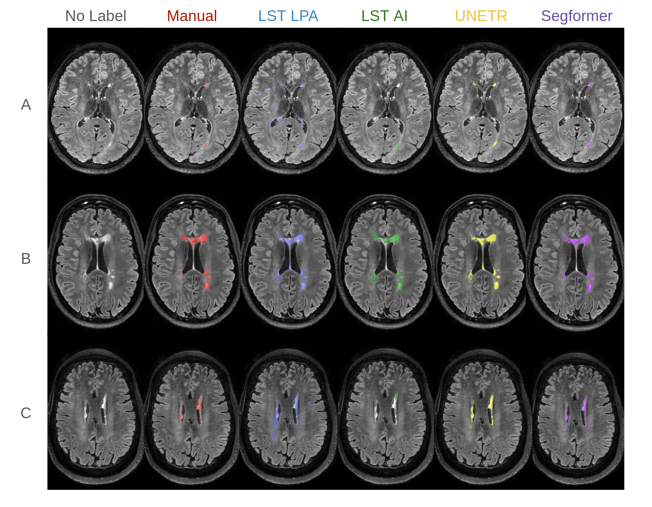
Figure 1: Comparative qualitative outcomes across segmentation methods at native 7T FLAIR resolution, highlighting differences in sensitivity to lesions and failure modes such as oversegmentation and missed small lesions.
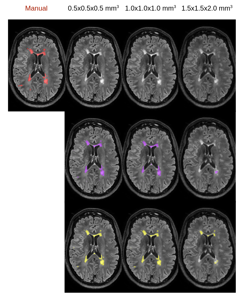
Figure 2: Effect of training image resolution on model performance, demonstrating loss of lesion detail and segmentation completeness at lower resolutions.
Artifact Handling and Failure Modes
Qualitative analysis with visual inspection reveals characteristic failure patterns. LST-LPA is prone to systematic oversegmentation, frequently labeling non-lesional hyperintense areas (e.g., bright cortical boundaries, choroid plexus) as WMLs. LST-AI reduces these spurious calls but underrepresents lesion volume and occasionally omits subtle foci, especially those with 7T-specific contrast profiles.
Transformer-based models (especially SegFormer) more closely recapitulate expert annotations, successfully capturing small and irregular lesions that are often omitted by classical methods. Furthermore, SegFormer demonstrates advantageous artifact resistance, notably avoiding false positive identification of wraparound artifacts, which confound both classical and other neural architectures.
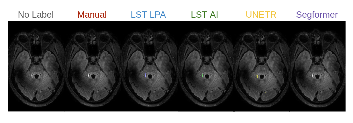
Figure 3: Differential model responses to 7T-specific artifacts, with SegFormer minimizing false positives compared to other approaches.
Methodological and Practical Implications
The study rigorously demonstrates that domain adaptation to 7T data is essential—models trained exclusively on legacy lower-field MRI (1.5T/3T) generalize poorly to 7T, failing to exploit its increased spatial resolution and suffering from domain shift. Transformer-based models trained on 7T data emerge as high-performing alternatives, especially for small-lesion sensitivity.
The superiority of high-resolution training is empirically validated, highlighting that ultra-high field imaging's incremental value is only realized when exploited by resolution-matched, context-aware learning frameworks. Moreover, by providing pretrained, reproducible models for the community, the work lowers technical entry barriers for standardized, automated WML quantification in centers adopting 7T MRI, advancing reproducibility and comparability in MS research.
However, performance rankings must be contextualized within the labeling pipeline, as use of LST-AI for some test mask initialization can skew overlap-based metrics in its favor, positioning its reported scores as optimistic upper bounds rather than fully independent benchmarks.
Future Research Directions
Several avenues warrant further exploration:
- Multicenter, cross-protocol generalization (robustness beyond a single scanner/institution)
- Integration of self-supervised, cross-modal, or large-scale pretraining strategies
- Modeling of additional lesion subtypes (e.g., gray matter lesions, cortical demyelination)
- Joint segmentation leveraging multi-contrast 7T data (T1w, T2w, FLAIR)
- Domain adaptation and federated learning frameworks for harmonization across institutions
Advancements in these dimensions will further close the gap between rapidly evolving hardware capabilities and the effective clinical/biomarker utility of high-field MRI.
Conclusion
This work establishes transformer-based architectures, when carefully adapted and trained with native 7T FLAIR data, as strong alternatives for automated MS lesion segmentation, offering substantial improvements over classical pipelines, especially for small lesion and artifact-prone regions. Importantly, small-lesion sensitivity is maximized only when models are exposed to native high-resolution data during training. Release of these models facilitates reproducible ultra-high field research pipelines and opens the path for further advances in multi-centric and multi-contrast analysis frameworks for MS.