- The paper demonstrates that engineered 2D split-Gaussian PSF and Bayesian inference allow sub-wavelength resolution (39 nm RMSE) without nonlinear saturation.
- It applies spatial mode demultiplexing in a TIRF microscope with a quadrant phase plate, effectively overcoming Rayleigh's curse for closely spaced emitters.
- Experimental results show a 1.6-fold improvement over conventional imaging, with resolutions closely matching the predicted Cramér-Rao bounds.
Structured Detection Microscopy: Enabling Far Sub-Wavelength Resolution without Nonlinear Saturation or Photoswitching
Introduction
"Structured Detection Microscopy" (2604.00413) presents a principled extension of spatial mode demultiplexing (SPADE) to biological imaging, delivering far sub-wavelength (down to 39 nm resolution for 50 nm emitter separations) measurement without reliance on nonlinear saturation or stochastic fluorophore switching. The work systematically addresses the limitations of established super-resolution techniques—such as STED, PALM/STORM, and SIM—by leveraging PSF engineering and Bayesian statistical inference to overcome Rayleigh's curse, maintaining compatibility with standard camera-based detection, and integrating seamlessly with high-NA TIRF fluorescence microscopy platforms.
The foundational concept is the restructuring of the image plane PSF to decouple signal information from high shot-noise regions, thus boosting the Fisher information for closely spaced emitters. Unlike previous SPADE demonstrations, which were restricted to 1D or used mode-sorting optics incompatible with camera arrays, the authors engineer a 2D 'split-Gaussian' PSF. This mode (approximating a TEM1,1 Hermite-Gauss profile) enables enhanced localization precision for point emitters well below the diffraction limit.
The analysis reveals a separation-dependent regime: for emitter separations s<0.45σ (with σ the diffraction-limited width), the engineered PSF yields higher Fisher information (FI) than the conventional Gaussian PSF, with the Fisher information ratio diverging as s→0. This spatial structuring of detection modes is robust to emitter orientation, resulting in negligible FI variation across angles.
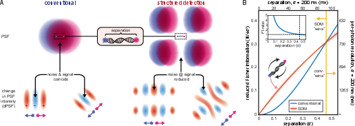
Figure 1: Theoretical Fisher information and its spatial distribution for conventional versus structured detection, demonstrating the SDM advantage for sub-diffraction separations.
Experimental Implementation: Structured Detection in a TIRF Microscope
The system is realized by integrating a quadrant phase plate into the Fourier plane of a commercial 3i Marianas TIRF microscope. This phase plate introduces a π phase shift in diagonally opposed quadrants, generating the split-Gaussian PSF upon detection while maintaining optical throughput compatible with an EMCCD camera (iXon Ultra 897). DNA nanorulers (GATTA-quant) with separations of 50, 120, and 180 nm are labeled with Alexa Fluor 488 at each end and immobilized for imaging. The excitation is provided at 488 nm, with fluorescence collected at 519 nm and isolated from excitation and background via a cascade of bandpass and dichroic filters.
Photobleaching, ambient, and electronic noise are systematically minimized, and the EMCCD is operated at low temperature and high gain for maximal signal fidelity.
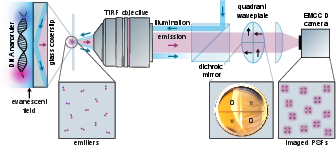
Figure 2: Schematic of the SDM experimental setup, illustrating TIRF excitation, phase plate insertion in the Fourier plane, and image formation pathways.
Bayesian Data Analysis Pipeline
The separation estimation is treated as a probabilistic inference problem. For each imaged emitter pair, the photon detection events serve as the dataset, with their spatial distribution modeled by appropriate split-Gaussian or Gaussian PSF templates. The initial parameter space comprises seven dimensions (positions, PSF widths, noise), but this is reduced to two (separations in x and y) by direct centroid evaluation and empirical noise modeling, making Bayesian computation tractable.
The posterior over emitter separations is constructed by multiplying per-detection likelihood functions, normalized over a finely sampled grid. Corrective bias functions, calibrated for each imaging modality and separation regime via simulation under matching SNR and count statistics, are applied to the maximum a posteriori estimates.
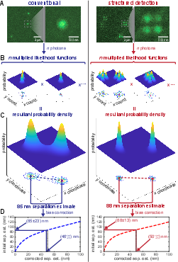
Figure 3: Data analysis pipeline, from PSF imaging to likelihood construction, posterior computation, and bias correction for separation estimation.
Results: Achieved Resolution and Statistical Validation
Empirical validation over multiple nanoruler lengths (N=50, 120, 180 nm) demonstrates that SDM consistently yields superior localization precision compared to conventional imaging. For 50 nm rulers, SDM achieves 39 nm RMSE versus 64 nm for conventional, a 1.6-fold improvement, with similar enhancements for larger separations. The experimental resolutions are in close agreement with predicted Cramér-Rao bounds, with minor deviations attributable to nanoruler length dispersity, detector nonidealities, and residual model/systematic errors.
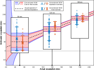
Figure 4: Experimental resolutions and means for SDM versus conventional microscopy across nanoruler separations, with Cramér-Rao bounds overlaid.
Numerical FI/CRB analysis, informed by the actual image statistics, corroborates the empirical trend that the theoretical resolution advantage of SDM emerges for separations well below the diffraction limit and is, in practice, only slightly mitigated by real-world noise and pixelation.
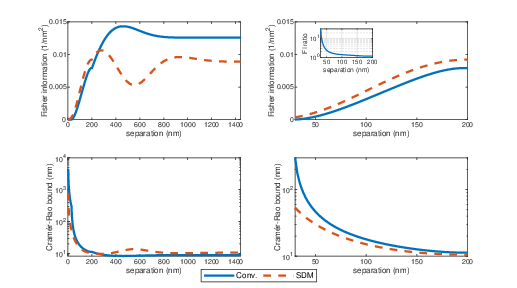
Figure 5: Simulated Fisher information and Cramér-Rao bound profiles, with experimental data overlay. The SDM CRB remains below the conventional CRB across the relevant parameter regime.
Discussion: Implications, Limitations, and Outlook
SDM, as established here, is capable of extracting inter-emitter separations at fivefold below the diffraction barrier, rivaling established techniques (e.g., PALM, STORM, STED) in achievable precision while operating with substantially lower photon dose and without the requirement for saturation or stochastics. This has unique implications for low-phototoxicity imaging and will enable super-resolution of systems inaccessible to conventional methods, e.g., intrinsically luminescent biomolecular assemblies.
Practical integration within a TIRF system underscores immediate applicability. However, the current SDM protocol is fundamentally limited to two-emitter separation estimation. Generalization to higher-order or arbitrary incoherent source distributions, necessary for broad biological imaging, will require adaptive optical elements (e.g., programmable phase arrays) or sequential mode projections, building on the work in advanced SPADE and adaptive optics [Matlin:2022]. Real-time data processing is currently a computational bottleneck but is amenable to optimization and hardware acceleration.
Advances in noise suppression, improved quantum efficiency, and increased photon budget (by mitigating photobleaching or extending acquisition) can push SDM further—modeling suggests 5 nm resolution is within reach under optimal conditions.
Conclusion
Structured Detection Microscopy delivers experimentally validated far-sub-wavelength resolution in a camera-based microscopy architecture without the photon budget or sample preparation requirements of nonlinear or stochastic methods. It robustly overcomes Rayleigh's curse via spatial PSF engineering and principled Bayesian inference. While limited to pairwise separation analysis in its current form, the demonstrated architecture is poised for extension to complex source distributions and broader live-cell biomicroscopy applications. Future technical advances in adaptive optics and computation will further increase its relevance for biological and quantum imaging (2604.00413).