Multiscale Causal Geometric Deep Learning for Modeling Brain Structure
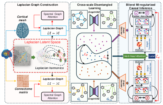
Abstract: Multimodal MRI offers complementary multi-scale information to characterize the brain structure. However, it remains challenging to effectively integrate multimodal MRI while achieving neuroscience interpretability. Here we propose to use Laplacian harmonics and spectral graph theory for multimodal alignment and multiscale integration. Based on the cortical mesh and connectome matrix that offer multi-scale representations, we devise Laplacian operators and spectral graph attentions to construct a shared latent space for model alignment. Next, we employ a disentangled learning combined with Graph Variational Autoencoder architectures to separate scale-specific and shared features. Lastly, we design a mutual information-informed bilevel regularizer to separate causal and non-causal factors based on the disentangled features, achieving robust model performance with enhanced interpretability. Our model outperforms baselines and other state-of-the-art models. The ablation studies confirmed the effectiveness of the proposed modules. Our model promises to offer a robust and interpretable framework for multi-scale brain structure analysis.
Paper Prompts
Sign up for free to create and run prompts on this paper using GPT-5.
Top Community Prompts
Explain it Like I'm 14
What is this paper about?
This paper is about building a smart computer model that can understand the brain’s structure using two types of MRI scans. It combines:
- Structural MRI (sMRI), which shows the shape and thickness of the brain’s outer layer (like the hills and valleys of the brain surface).
- Diffusion MRI (dMRI), which shows how different brain areas are connected by white matter pathways (like roads between neighborhoods).
The main goal is to merge these two kinds of information, across different scales, in a way that is both accurate and easy for neuroscientists to interpret.
What questions did the researchers ask?
The researchers wanted to find simple answers to these key questions:
- How can we combine the “shape” of the brain (from sMRI) and the “connections” in the brain (from dMRI) so they work together?
- Can we build a method that separates what is shared between the scans and what is unique to each scan?
- Can we figure out which features truly cause good predictions (like age or gender) and which features are just noise?
How did they study it?
Think of the brain like a city:
- The brain’s surface (from sMRI) is like the city’s map: hills, roads, and neighborhoods.
- The brain’s connections (from dMRI) are like the highways and streets linking different places.
The researchers created a three-part system to learn from both “maps” at once.
1) Creating a shared “wave” space (Laplacian Latent Space)
- They turned both the brain surface and brain connections into something like musical waves called “Laplacian harmonics.”
- These waves can be long (capturing big, global patterns across the brain) or short (capturing small, local details). Combining waves of different lengths helps the model understand the brain at multiple scales.
- They aligned the waves from the two data types into the same shared space, so the shape and connection information could “talk” to each other.
- They used a method called Spectral Graph Attention, which is like teaching the model to “listen” more closely to the most important connections between brain areas while keeping the wave patterns intact.
2) Separating shared and unique features (Cross-scale Disentangled Learning)
- Even in the shared space, sMRI and dMRI have different strengths. The model separates:
- Shared features (what both scans agree on), and
- Unique features (what is special to each scan).
- They used a Graph Variational Autoencoder (GraphVAE), which is like a translator: it tries to rebuild dMRI using information from sMRI, and rebuild sMRI using information from dMRI. If the translation works well, the model has learned meaningful shared features.
- This “disentangling” helps avoid mixing unrelated signals and makes the results more structured and easier to interpret.
3) Finding likely causes, not just correlations (Bilevel MI-regularized Causal Inference)
- The model tries to separate features into:
- Causal features: signals that truly help predict age or gender.
- Non-causal features: signals that look related but are just noise or coincidences.
- It uses mutual information (MI), which measures “how much knowing one thing tells you about another,” to encourage the model to keep useful, predictive features and reduce spurious ones.
- It also looks at how sMRI and dMRI causal features interact across modalities—like checking if both the city map and the highway map point to the same important places.
- Finally, it feeds the cleaned-up features into a graph transformer (a powerful deep learning tool for graphs) to make final predictions.
What did they find and why it matters?
The researchers tested their model on 115 participants from the Human Connectome Project (HCP). They tried predicting:
- Age (a number)
- Gender (a category)
Here’s what stood out:
- The new model beat other methods in predicting age, with a lower error (MAE ≈ 2.36 years; RMSE ≈ 2.90). That means it’s more precise.
- It also did well in gender prediction (about 79% accuracy), better than most competing models.
- When they removed parts of the system (in ablation studies), performance dropped. The biggest drop happened when they removed the disentangling module—showing that separating shared and unique information is crucial.
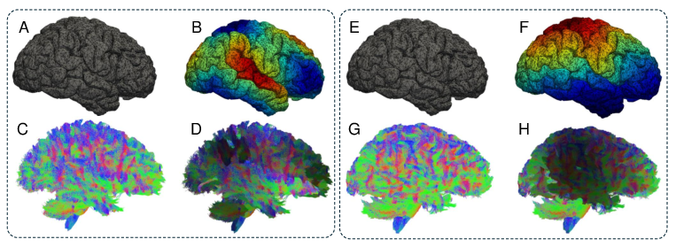
- They visualized “wave heatmaps” and found that younger and older participants had different patterns in both the brain surface and connections, which supports the idea that harmonics capture meaningful brain differences.
This matters because:
- It shows a way to combine different brain data types that is both strong and scientifically interpretable.
- It reduces the risk of the model learning “fake” patterns and increases trust in the results.
What could this mean in the real world?
- Better tools for studying brain development and aging: The model can find patterns across scales that relate to age and possibly to diseases like Alzheimer’s.
- More reliable and interpretable biomarkers: Because the method focuses on causal features, doctors and scientists can have more confidence in what the model finds.
- Stronger multimodal analysis: Many brain conditions affect both structure and connections, and this framework can capture both.
- Future directions: With more data and testing on clinical populations, this approach could support diagnosis, track disease progression, and help personalize treatments by revealing meaningful multiscale brain patterns.
Knowledge Gaps
Knowledge gaps, limitations, and open questions
Below is a single, focused list of concrete gaps and unresolved questions that future work could address to strengthen the paper’s claims and utility.
- External validity and cohort diversity: Results are based on the first 115 HCP subjects (largely young adults) with no external validation. How does performance generalize across age spans, scanners, sites, and clinical populations (e.g., AD, MCI, glioma, psychiatric cohorts like ADNI, UK Biobank, ABCD)?
- Sample size and statistical rigor: With N=115, model complexity risks overfitting. Are gains statistically significant (e.g., permutation tests, confidence intervals, effect sizes)? How stable are results across multiple random splits and seeds?
- Assumption of mesh–connectome one-to-one mapping: The paper assumes each mesh vertex corresponds to a connectome node, which is atypical (connectomes are usually region-based). What parcellation/resolution is used, and how sensitive are results to parcellation choice (Desikan, Schaefer), mesh resolution, and node definition?
- Connectome construction details: The connectome Laplacian depends on tractography choices (iFOD2, ACT, number of streamlines, SIFT/SIFT2, thresholding, weight type). Which exact pipeline and parameters are used, and how do they affect the Laplacian spectrum and downstream performance?
- Fused Laplacian definition: The construction of the fused Laplacian is underspecified. What is its exact formulation (e.g., weighted sum, joint optimization, co-regularized eigenproblem, co-Laplacian)? What guarantees exist for spectral alignment and stability?
- Spectral alignment procedure: The Rayleigh quotient–based projection is described but not fully specified. How are eigenvectors/harmonics ordered, normalized, and disambiguated (sign/phase)? Is the alignment robust across subjects and to numerical instabilities?
- Spectral Graph Attention “preservation” claims: The paper claims attention preserves spectral properties, but standard GATs do not guarantee eigen-structure preservation. Can the authors provide mathematical guarantees or empirical checks (e.g., tracking eigenvalue/eigenvector drift across layers)?
- Graph construction via cosine similarity: Building edges from cosine similarity of dominant eigenvectors can be sensitive to noise and scaling. What thresholds, kNN settings, or normalization schemes are used? How sensitive are results to these design choices and to the number of harmonics ?
- Shapes and notation clarity: Key variables (N, F, k, d), feature tensors, and adjacency sizes are inconsistently defined. Can the authors specify exact tensor shapes, data dimensions, and the mapping between harmonics, nodes, and features for reproducibility?
- Disentangled learning identifiability: The split into “shared” and “unique” factors lacks identifiability guarantees. Under what conditions is disentanglement recoverable? How much leakage occurs between shared and unique subspaces (quantified via controlled synthetic benchmarks)?
- Disentangled loss specification: The loss in Eq. (6) is malformed and variables (e.g., ) are not fully defined. What is the exact expression, weighting, and optimization protocol? How does the union get computed?
- GraphVAE cross-reconstruction details: What reconstruction losses, priors, and KL annealing schedules are used? How do these design choices affect the alignment of shared spaces and the risk of memorization or mode collapse?
- Mutual information estimation: The bilevel MI loss requires differentiable MI estimators (e.g., MINE, NWJ, InfoNCE, kNN-MI). Which estimator is used, how is it stabilized, and what are its bias/variance properties on small samples?
- Sign of MI regularizers: The causal loss combines negative and positive MI terms. Minimizing the current objective may inadvertently reduce cross-modality MI (), contrary to the stated goal. Can the authors clarify the signs, optimization objective, and intended effect on cross-modality interactions?
- Causal validity: Without interventions, temporal data, or strong causal assumptions, the “causal vs. non-causal” feature split may be correlational. Can the authors validate causal claims via simulated datasets with known ground-truth causal structure, confounding controls, or causal discovery benchmarks?
- Neuroscience interpretability: Beyond heatmaps, are the learned harmonics and causal graphs reproducible across subjects and aligned with known neuroanatomy? Can the authors provide region-level analyses, enrichment tests, or stability maps relating spectral components to biology?
- Robustness and uncertainty: No analyses address registration errors, motion artifacts, noise perturbations, or missing modality scenarios. How robust are outputs to these factors, and can predictive uncertainty and calibration be reported?
- Hyperparameter sensitivity: Key hyperparameters (number of harmonics F, attention heads, , , learning rate, batch size) are not systematically explored. What is the sensitivity landscape, and how was tuning performed to avoid overfitting?
- Computational scalability: The approach may scale poorly with high-resolution meshes. What are memory/time complexities in terms of vertices K and harmonics F? Is multi-GPU training or approximation (e.g., subsampling, Nyström methods) needed for full-resolution cortical meshes?
- Task selection and clinical relevance: Only age and gender are evaluated. Can the framework be tested on clinically meaningful endpoints (diagnosis/prognosis in AD/MCI/glioma, cognitive scores) to support claims of pathology modeling?
- Fairness and bias: Gender classification shows high variability (F1 std ~0.29). Are there performance disparities across sex, age bins, handedness, or other demographics? Provide cohort statistics and fairness analyses.
- Baseline coverage and tuning fairness: Comparisons exclude some recent geometric and causal GNNs, and tuning protocols for baselines are unclear. Are all baselines given equivalent hyperparameter searches and data preprocessing?
- Evaluation reporting: Report calibration curves, reliability diagrams, and confidence intervals for metrics. Consider permutation tests and bootstrap confidence intervals to substantiate improvements.
- Positional encoding (): How are spatial distances computed (geodesic vs. Euclidean vs. anatomical distances), and how sensitive is attention to the choice of positional encoding?
- Cross-modality fusion design: Current fusion uses attention between modality outputs plus shared features. Would a multiplex or co-Laplacian approach, or coupled eigenproblems, yield better cross-scale integration? Comparative studies could clarify.
- Edge-case numerical stability: How are negative/zero eigenvalues, disconnected graphs, singular Laplacians, or degenerate spectra handled? Are regularizations (e.g., diagonal loading) applied to prevent over-smoothing or numerical issues?
- Reproducibility and availability: Code, exact preprocessing versions (FreeSurfer, MRtrix3 settings), random seeds, architectural details, and data splits are not provided. Making these available is critical for replication.
- Figure-level evidence: Heatmap differences are illustrative but anecdotal. A quantitative analysis linking specific harmonics (wavelength bands) to age/gender, with statistical tests, is needed to substantiate interpretability claims.
- Extension to additional modalities: How would the framework integrate fMRI, T2-FLAIR, or quantitative MRI (e.g., myelin maps), and how would the fused Laplacian and disentanglement scale with more modalities?
- Theoretical grounding: Provide conditions under which spectral alignment yields a valid shared latent space across modalities and subjects, and relate these to known results in spectral geometry and functional Laplacian harmonics.
Practical Applications
Immediate Applications
Below are actionable use cases that can be deployed now, leveraging the paper’s methods (Laplacian latent space alignment, spectral graph attention, cross-scale disentanglement with GraphVAE, and bilevel MI-regularized causal inference), along with sector links and feasibility notes.
- Multimodal neuroimaging analysis pipeline for research labs (academia; healthcare R&D)
- Use La-MuSe to integrate sMRI-derived cortical meshes with dMRI-derived connectomes for biomarker discovery, beyond age/sex (e.g., pilot analyses for MCI, TBI, developmental studies).
- Workflow: FreeSurfer + MRtrix3 preprocessing → Laplacian matrices → spectral graph attention refinement → disentangled shared/unique features → bilevel causal modeling → visualization (harmonic maps and causal graphs).
- Potential tools/products: A PyTorch/PyG-based open-source codebase; neuroimaging visualization modules for harmonics and causal graphs; QC dashboards to flag spurious connections.
- Dependencies/assumptions: Accurate coregistration and parcellation; high-quality tractography; harmonics well-aligned via Rayleigh quotient; stable MI estimation; sufficient sample sizes for generalization.
- Retrospective risk stratification studies (healthcare; hospital analytics)
- Apply the pipeline to existing institutional datasets to explore multiscale brain structural signatures associated with conditions (e.g., early neurodegeneration signals; white matter injury patterns).
- Potential workflow: Reprocess historical sMRI/dMRI cohorts → compute multiscale features → correlate with outcomes; report interpretable causal graphs for clinical investigators.
- Dependencies/assumptions: IRB/ethics approvals; robust data harmonization across scanners and protocols; domain alignment of mesh vertices to connectome nodes.
- Connectome-aware presurgical research models (neurosurgery research)
- Build prototypes for predicting deficits (e.g., language decline) using multiscale features, complementing prior connectome work cited.
- Potential tools/products: Research-only decision support dashboards showing causal subgraphs linked to predicted risk, to inform study design and hypothesis generation.
- Dependencies/assumptions: Requires patient cohorts with surgical outcomes; method currently validated on healthy subjects; not yet clinical-grade.
- Cohort selection and enrichment for clinical trials (pharma/biotech)
- Use “brain age” deviation and multiscale structure/cause graphs to select or enrich trial populations (e.g., early converters, responders).
- Workflow: Centralized imaging processing → La-MuSe feature extraction → stratification rules; incorporate interpretable reports into trial operations.
- Dependencies/assumptions: Standardized imaging protocols; regulatory alignment for trial biomarker usage; reproducibility across sites.
- Cross-modal alignment toolkit for other brain modalities (academia; software tools)
- Adapt the Laplacian latent space + spectral graph attention to align sMRI+dMRI with fMRI or MEG/EEG-derived network graphs (research prototypes).
- Potential tools/products: “Spectral Alignment” library; plug-ins for MNE, Nilearn, or BrainSuite.
- Dependencies/assumptions: Requires compatible parcellations; careful treatment of temporal vs structural scales.
- Curriculum and teaching modules on spectral geometric deep learning in neuroimaging (education)
- Develop lab exercises demonstrating Laplacian harmonics, eigenvector alignment, graph attention, and causal inference on HCP subsets.
- Potential tools/products: Course notebooks, interactive harmonic heatmap viewers.
- Dependencies/assumptions: Access to curated datasets; computational resources similar to a single GPU workstation.
- Method transfer to other multimodal geometric graph problems (software/engineering)
- Immediate prototypes in non-medical domains with geometry+network data (e.g., civil infrastructure: mesh of structures + sensor networks; materials: microstructure meshes + lattice connectivity).
- Potential tools/products: Internal R&D SDK applying La-MuSe’s alignment/disentanglement/causal modules to industrial graph-geometry data.
- Dependencies/assumptions: Availability of paired geometric and network representations; domain-specific validation of causal interpretations.
Long-Term Applications
These opportunities require further research, scaling, clinical validation, or productization, building on the framework’s interpretability and performance.
- Regulatory-grade diagnostic decision support for brain disorders (healthcare; medical devices)
- Use multiscale harmonics and causal graphs as interpretable biomarkers for AD/MCI, MS, TBI, psychiatric conditions.
- Potential products: PACS-integrated modules; clinician-facing reports with causal subgraph explanations; model monitoring for drift.
- Dependencies/assumptions: Large multi-site validation; bias/fairness assessments; standardized pipelines; regulatory approval; robust calibration across scanners.
- Personalized neurosurgical planning and simulation (healthcare; surgical informatics)
- Predict functional deficits and simulate resection effects using patient-specific multiscale structural causal graphs; integrate with intraoperative mapping tools.
- Potential products: Preoperative planning platforms; digital twin of patient brain structure-function relationships.
- Dependencies/assumptions: Prospective clinical studies; integration with functional data; uncertainty quantification; surgeon workflow integration.
- Closed-loop neurostimulation targeting via structural causal graphs (healthcare; neurotech)
- Use inferred causal subgraphs to design stimulation targets (DBS/TMS); combine with fMRI/EEG for closed-loop control.
- Potential products: Decision engines interfacing with neurostimulation devices; adaptive stimulation protocols informed by multiscale features.
- Dependencies/assumptions: Strong causal validity; safety and efficacy trials; multimodal integration; device interfaces and standards.
- Longitudinal brain health monitoring (consumer health; population health)
- Track “brain age” trajectories and multiscale structural changes over time to detect early deviations; inform preventive interventions.
- Potential products: Clinical monitoring platforms; population-level dashboards; insurers/payers analytics.
- Dependencies/assumptions: Secure data pipelines; privacy-preserving analytics; normative atlases; evidence linking trajectories to outcomes.
- Cloud-based, multi-institution imaging AI platform for multiscale causal modeling (software; healthcare IT)
- Offer standardized preprocessing, model inference, and interpretability reporting at scale; support cross-site studies and registries.
- Potential products: Managed service; API-accessible La-MuSe; auditability tools for causal graphs.
- Dependencies/assumptions: Interoperability with hospital IT; data governance; reliability and uptime; cost-effective GPU scaling.
- Surrogate endpoints and responder prediction in drug development (pharma)
- Validate multiscale harmonics/causal features as surrogate endpoints; predict responders based on structural network signatures.
- Potential products: Biomarker qualification dossiers; companion analytics aligned with trial endpoints.
- Dependencies/assumptions: Regulatory biomarker qualification; harmonized imaging across trials; causal interpretability accepted by regulators.
- Full multimodal brain integration beyond sMRI/dMRI (academia; healthcare R&D)
- Extend to fMRI, MEG/EEG, and even histology/genomics (multi-omics) using cross-scale disentanglement and causal regularization for integrative brain models.
- Potential products: Comprehensive atlas of structure-function-cause relationships; research platforms for translational studies.
- Dependencies/assumptions: Cross-domain alignment challenges; time-frequency vs spatial scales; large, curated multimodal datasets.
- General-purpose Spectral Graph AI SDK for multiscale geometric data (software; robotics; autonomous systems; energy)
- Productize core ideas—Laplacian latent space alignment, spectral attention, disentangled cross-reconstruction, and MI-regularized causal inference—for domains such as:
- Robotics/autonomy: city mesh + traffic graphs; map-building with interpretable causal connectivity.
- Energy: grid topology + physical infrastructure meshes; causal reliability analysis.
- Manufacturing/materials: microstructure meshes + connectivity graphs; interpretable defect causality.
- Potential products: Enterprise SDK; consultancy packages; benchmarking suites.
- Dependencies/assumptions: Domain adaptation; appropriate definitions of “causal” in physical systems; validation against ground truth.
- Policy and standards development for interpretable multimodal imaging AI (policy; standards bodies)
- Establish best practices for alignment, harmonics-based features, and reporting of causal graphs; guidelines for dataset curation and privacy.
- Potential outputs: Standard operating procedures; interpretability report templates; governance frameworks.
- Dependencies/assumptions: Multi-stakeholder coordination; empirical evidence; alignment with regulatory expectations.
Cross-cutting assumptions and dependencies
- Data quality and harmonization: Accurate coregistration (mesh–connectome), consistent parcellations, tractography reliability, and site/scanner harmonization are crucial.
- Model stability: Mutual information estimation and disentanglement need careful tuning and may be sensitive to sample size and noise.
- Generalization and fairness: Current validation (115 HCP subjects, age/sex) is limited; larger, diverse cohorts are required to assess biases and robustness.
- Interpretability: Causal graphs are model-derived; causal claims require domain validation, counterfactual testing, or interventional evidence in clinical contexts.
- Compliance: Clinical deployments require regulatory approval, privacy/security controls, and alignment with clinical workflows.
Glossary
- 5-tissue-type (5tt) segmentation: A tissue-class segmentation that produces five tissue labels used to constrain tractography. "Then, we used 5-tissue-type (5tt) segmentation with Freesurfer outputs to generate a gray matter-white matter interface."
- Anatomically-Constrained Tractography (ACT): A diffusion MRI tractography method that restricts fiber tracking using anatomical priors from tissue segmentation. "Finally, we performed fiber tracking using the iFOD2 algorithm with MRTRIX3's ACT function."
- Bilevel MI-regularized Causal Inference: A causal modeling module that uses mutual information to separate causal from non-causal features within and across modalities. "we propose a Bilevel Mutual-information (MI)-regularized Causal Inference Module modelling cross-modality interactions and modality-specific causal graphs."
- Connectome matrix: A matrix representation of structural brain connectivity between regions, typically derived from diffusion MRI tractography. "we input the cortical mesh derived from T1-weighted MRI and connectome matrix derived from dMRI into the Laplacian latent space for modality alignment."
- Coregistration: The process of aligning images or modalities into a common coordinate space so that corresponding anatomical locations match. "high-precision cortical meshes and connectome matrices are coregistered so that each vertex on the mesh corresponds to a node in the connectome."
- Cortical mesh: A triangular surface representation of the cerebral cortex used to model macrostructural morphology. "Firstly, we input the cortical mesh derived from T1-weighted MRI and connectome matrix derived from dMRI into the Laplacian latent space for modality alignment."
- Cotangent weights: Discrete mesh Laplacian weights computed from the cotangents of angles opposite mesh edges, used to approximate the Laplacian operator on surfaces. "The Laplacian matrix, , is constructed based on the cotangent weights: is calculated by "
- Cross-scale Disentangled Learning: A learning strategy that separates modality-specific and shared latent factors across scales to improve interpretability and fusion. "we devise a Cross-scale Disentangled Learning module to disentangle shared and unique features."
- Diffusion MRI (dMRI): An MRI modality that measures water diffusion to infer microstructural properties of white matter and connectivity. "diffusion MRI (dMRI) maps white matter microstructure."
- Disentangled features: Latent representations partitioned into distinct factors (e.g., causal vs. non-causal) to reduce confounding and improve interpretability. "Within each modality, the disentangled features $\mathbf{Z}^{\text{uni}$ are decomposed into causal () and non-causal () features:"
- FreeSurfer 'recon-all' pipeline: A standardized neuroimaging workflow for cortical surface reconstruction and morphometric analysis from T1-weighted MRI. "The T1 images were processed using Freesurfer's 'recon-all' pipeline before extracting cortical meshes"
- Graph transformer: A transformer architecture adapted to graph-structured data, using attention over nodes/edges with graph-specific positional encodings. "The final features are fed into graph transformers for downstream predictions."
- Graph Variational Autoencoder (GraphVAE): A variational autoencoder specialized for graph data that learns latent representations enabling graph reconstruction or generation. "we devise a dual-branch GraphVAE architecture"
- HCP dataset: A large-scale neuroimaging dataset from the Human Connectome Project, widely used for studying brain connectivity and structure. "We included the first 115 subjects from the HCP datasets."
- iFOD2 algorithm: A second-order integration tractography algorithm used in diffusion MRI to probabilistically propagate streamlines. "Finally, we performed fiber tracking using the iFOD2 algorithm with MRTRIX3's ACT function."
- Laplacian harmonics: Eigenfunctions of the Laplacian operator on meshes or graphs that provide multiscale, frequency-domain representations. "At each vertex, we compute the Laplacian harmonics as follows:"
- Laplacian latent space: A shared representation space defined by the eigenvectors of a fused Laplacian used to align multimodal geometric data. "we input the cortical mesh derived from T1-weighted MRI and connectome matrix derived from dMRI into the Laplacian latent space for modality alignment."
- Laplacian operator: A differential operator (or its discrete counterpart on meshes/graphs) capturing curvature and smoothness; its spectrum encodes multiscale structure. "The Laplacian operator \cite{popov2022spectrum,pang2023geometric,anand2023hodge}, a fundamental mathematical tool, can capture high-frequency details and fine-grained structures"
- MNI152 space: A standardized brain template (average of 152 subjects) used for spatial normalization in neuroimaging. "Firstly, all T1 and dMRI were registered to the MNI152 space"
- MRTRIX3: An open-source software suite for diffusion MRI processing, tractography, and visualization. "The dMRI were processed by MRTRIX3 \cite{TOURNIER2019116137} with eddy current and geometric distortion correction."
- Mutual information (MI): An information-theoretic measure of statistical dependence between variables, used here to regularize causal feature learning. "inspired by MI \cite{zheng2024ci}, to enhance model reliability and interpretability, we propose a Bilevel Mutual-information (MI)-regularized Causal Inference Module"
- Rayleigh quotient: A scalar functional used to relate vectors and symmetric matrices, often to estimate eigenvalues or align eigenvectors. "The and are projected onto the latent space by the Rayleigh quotient"
- Spectral Graph Attention: An attention mechanism that refines graph edges while preserving spectral (eigen) properties of the Laplacian. "Spectral Graph Attention which dynamically adjusts edge weights while preserving spectral properties."
- Spectral positional encoding: Positional encodings derived from graph Laplacian eigenvectors (spectral basis) to inform attention mechanisms about spatial relationships. "multi-head graph attention with spectral positional encoding:"
- Voronoi area: The local surface area associated with a mesh vertex, used in discrete Laplacian formulations to weight contributions. " denotes the Voronoi area at vertex "
Collections
Sign up for free to add this paper to one or more collections.