Building a Synthetic Vascular Model: Evaluation in an Intracranial Aneurysms Detection Scenario
Published 4 Nov 2024 in cs.CV and cs.AI | (2411.02477v1)
Abstract: We hereby present a full synthetic model, able to mimic the various constituents of the cerebral vascular tree, including the cerebral arteries, bifurcations and intracranial aneurysms. This model intends to provide a substantial dataset of brain arteries which could be used by a 3D convolutional neural network to efficiently detect Intra-Cranial Aneurysms. The cerebral aneurysms most often occur on a particular structure of the vascular tree named the Circle of Willis. Various studies have been conducted to detect and monitor the aneurysms and those based on Deep Learning achieve the best performance. Specifically, in this work, we propose a full synthetic 3D model able to mimic the brain vasculature as acquired by Magnetic Resonance Angiography, Time Of Flight principle. Among the various MRI modalities, this latter allows for a good rendering of the blood vessels and is non-invasive. Our model has been designed to simultaneously mimic the arteries' geometry, the aneurysm shape, and the background noise. The vascular tree geometry is modeled thanks to an interpolation with 3D Spline functions, and the statistical properties of the background noise is collected from angiography acquisitions and reproduced within the model. In this work, we thoroughly describe the synthetic vasculature model, we build up a neural network designed for aneurysm segmentation and detection, finally, we carry out an in-depth evaluation of the performance gap gained thanks to the synthetic model data augmentation.
The paper demonstrates a synthetic 3D vascular model (VaMos) that augments TOF-MRA data to enhance intracranial aneurysm detection.
It details innovative methods including arterial geometry modeling, aneurysm simulation, and noise generation to mimic realistic brain vasculature.
The approach boosted lesion sensitivity to 88.97% in a 3D U-Net framework, despite a moderate rise in false positive rates.
Synthetic Vascular Model for Intracranial Aneurysm Detection
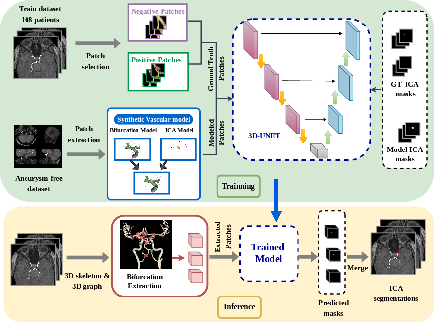
This paper introduces a novel approach to generating synthetic 3D vascular models for training deep learning algorithms, specifically for intracranial aneurysm (ICA) detection in Time-of-Flight Magnetic Resonance Angiography (TOF-MRA) images. The model aims to address the challenge of limited annotated data in medical imaging by creating a substantial dataset of realistic brain arteries, bifurcations, and ICAs. The effectiveness of this synthetic data augmentation is evaluated by training a 3D convolutional neural network for ICA segmentation and detection.
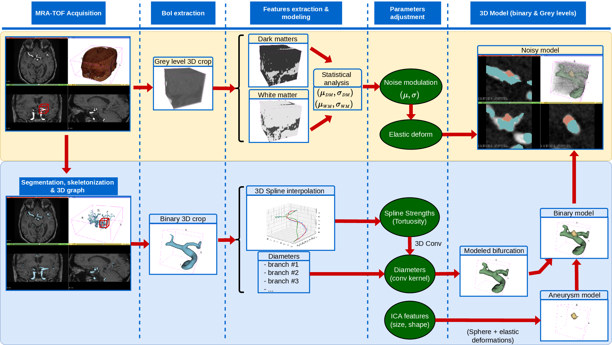
Methods: Synthetic Vascular Model (VaMos)
The synthetic vascular model, referred to as VaMos, comprises three main components: arterial geometry modeling, TOF noise simulation, and aneurysm generation (Figure 1).
Figure 1: Schematic representation of the whole bifurcation model, encompassing background noise modeling and arterial geometry modeling.
Arterial Geometry Modeling
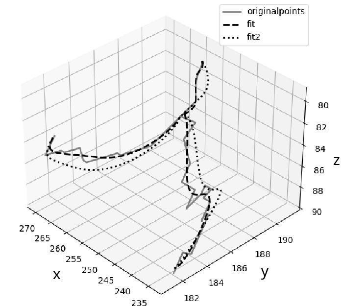
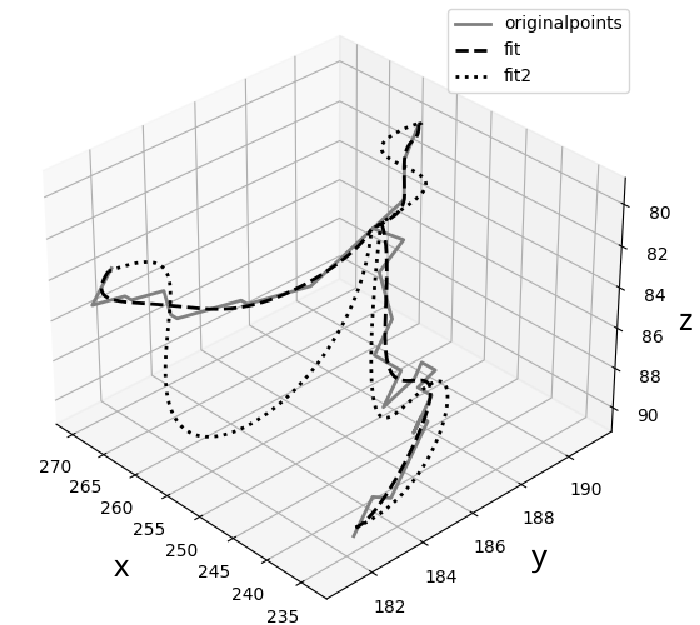
The arterial geometry is modeled by first extracting a 3D graph from segmented TOF volumes. Bifurcations of interest are automatically located, and 3D patches are cropped around the bifurcation nodes. Within these patches, the 3D coordinates of the arteries' centerlines are extracted and fitted using 3D spline functions. The spline functions are represented by knot-points, B-spline coefficients, and the order of the spline. Modifying the polynomial coefficients allows for the distortion of the centerline coordinates, mimicking the structural variability observed in human vasculature. The diameters of the arteries are then determined using a vascular tree characterization tool, and each centerline is convolved with a spherical kernel, adjusted to the corresponding diameter, to thicken the arteries. Elastic deformations are applied to the convolution kernel to avoid producing perfectly tubular arteries. Finally, a target gray level amplitude is set for the modeled vasculature. Examples of modified bifurcation centerlines are shown in (Figure 2).
Figure 2: Examples of modified bifurcation centerlines, showing the original branch, spline fit, and exaggerated modification of spline coefficients.
Aneurysm Modeling
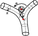
Synthetic aneurysms are incorporated into the model by first creating a 3D sphere and then distorting it using elastic deformations. The ICA center is aligned onto the bisector between the two daughter arteries, and the distance between the aneurysm center and the bifurcation node is computed based on the radii of the ICA and bifurcation branches, as well as the angle formed by the daughter arteries (Figure 3). A growth parameter is introduced to modulate the distance, allowing for the modeling of various states of growth for a given aneurysm.
Figure 3: Computation of the distance separating the aneurysm and the bifurcation center, with the ICA located along the bisector.
Background Noise Modeling
The background noise, representing the surrounding brain matter, fluids, and acquisition noise, is modeled by first separating the different brain components based on their radio-opacity. A multi-threshold segmentation technique is used to distinguish between hypointense and hyperintense matters, excluding the vasculature. Each matter is then geometrically distorted before generating and applying its overlaying noise. The noise generation process involves creating a high-frequency Gaussian noise of average set to the target 3D crop. This noise is then smoothed using a Gaussian filter with a standard deviation, σG, that is determined based on the target standard deviation, σf, of the filtered image, using the formula σG≈σ0/(2σfπ), where σ0 is the standard deviation of the input image. The resulting noise is then added to the geometric modeling of the bifurcation and aneurysm.
Model Assessment
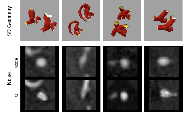
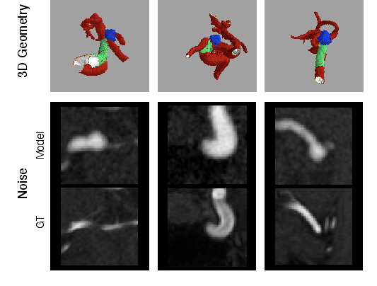
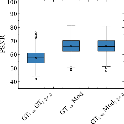
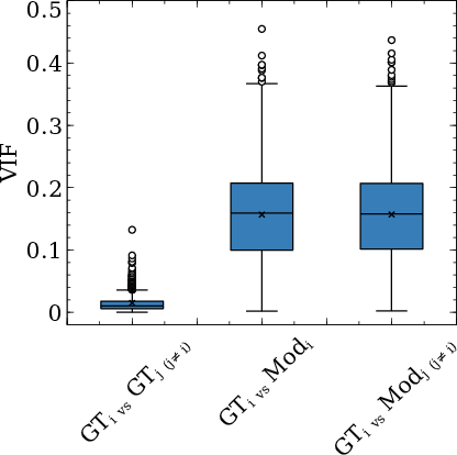
The authors assess the similarity of the synthetic and real images by comparing the modeled bifurcations with ground truth crops from TOF images (Figure 4). Both visual and quantitative assessments are performed. For the latter, several objective quality assessment metrics are used, including PSNR and VIF (Figure 5).
Figure 4: Comparison between modeled bifurcations and ground truth crops from a TOF, showing gray level voxel patches and 3D bifurcation layouts.
Figure 5: Objective assessment of the similarities between the ground truth patches and synthetic models, using PSNR and VIF metrics.
Experimental Design and Results
The synthetic model is used to generate a substantial training dataset for a 3D U-Net model, which is trained for ICA mask segmentation and detection. The dataset consisted of 190 TOF scans with unruptured ICAs, collected from multiple French institutions. A trained operator performed the annotations, and a neuroradiologist reviewed the cases. The overall aneurysm detection process is illustrated in (Figure 6).
Figure 6: Overview of the global procedure, encompassing the training step using synthetic images and the inference step.
Training and Evaluation
Three experiments were conducted to evaluate the effectiveness of the synthetic data augmentation:
Exp.#1: A baseline model trained using only real TOF patches.
Exp.#2: A model trained using a combination of real TOF patches and synthetic VaMos patches.
The models were evaluated using a four-fold cross-validation approach, and the performance metrics included lesion-level sensitivity and false positive rate (number of false positives per TOF).
Results
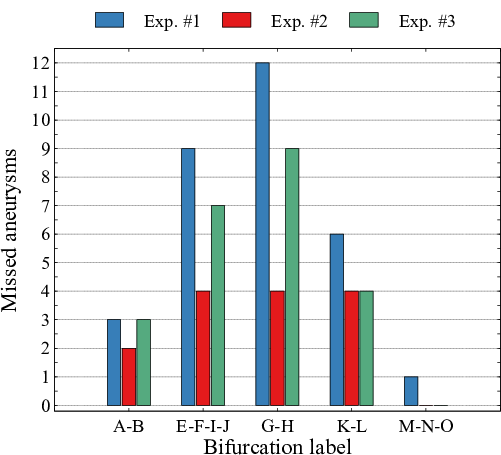
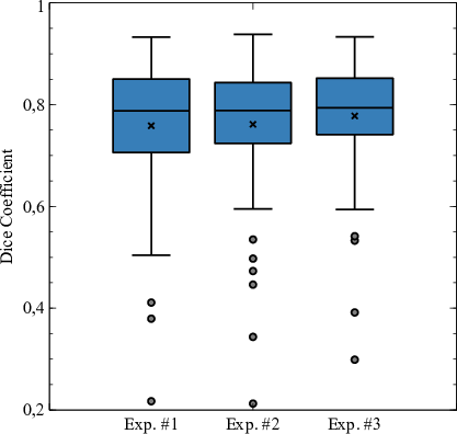
The results showed that Exp.#2, which used a combination of real and synthetic data, achieved the highest lesion-level sensitivity (88.97\%) compared to Exp.#1 (75.60\%) and Exp.#3 (81.88\%). However, Exp.#2 also had a higher false positive rate (0.40) compared to Exp.#1 (0.22) and Exp.#3 (0.36). The impact of aneurysm size and location on the detection performance was also analyzed, revealing that the synthetic data augmentation improved the detection rate for smaller aneurysms and reduced the number of missed aneurysms in specific locations along the Circle of Willis (Figure 7). The Dice scores for the detected aneurysms were comparable across the three experiments (Figure 8).
Figure 7: Missed detections with respect to the aneurysms positions in the test dataset for all three tested experiments.
Figure 8: Dice similarity coefficient of true ICAs for all three tested experiments.
Ablation studies were conducted to validate the various components of VaMos, including the noise distribution and the number of augmented patches, and the sensitivity for small aneurysms was greatly improved by the VaMos approach.
Discussion and Conclusion
The synthetic vasculature model effectively counterfeits TOF image portions, improving the performance of deep learning algorithms for ICA detection. The combination of real and synthetic data leads to a significant improvement in sensitivity, although it may also increase the false positive rate. The model's flexibility allows for adaptation to other imaging modalities, such as CTA or DSA. Further research is needed to explore the use of larger datasets and to refine the synthetic model parameters to further improve the detection performance for small aneurysms and to reduce the false positive rate.