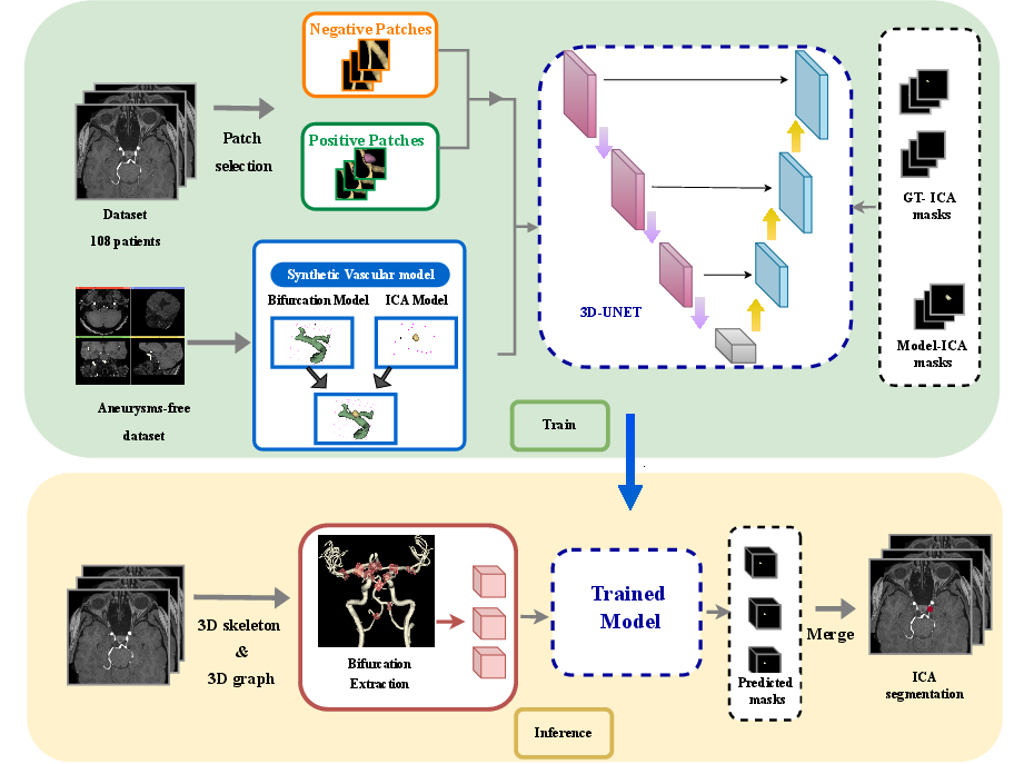
A vascular synthetic model for improved aneurysm segmentation and detection via Deep Neural Networks
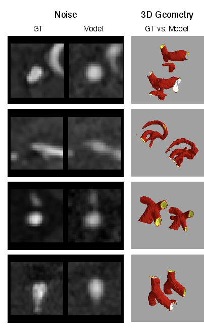
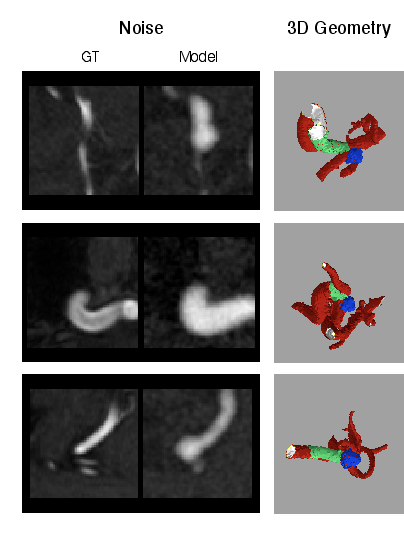
Abstract: We hereby present a full synthetic model, able to mimic the various constituents of the cerebral vascular tree: the cerebral arteries, the bifurcations and the intracranial aneurysms. By building this model, our goal was to provide a substantial dataset of brain arteries which could be used by a 3D Convolutional Neural Network (CNN) to either segment or detect/recognize various vascular diseases (such as artery dissection/thrombosis) or even some portions of the cerebral vasculature, such as the bifurcations or aneurysms. In this study, we will particularly focus on Intra-Cranial Aneurysm (ICA) detection and segmentation. The cerebral aneurysms most often occur on a particular structure of the vascular tree named the Circle of Willis. Various studies have been conducted to detect and monitor the ICAs and those based on Deep Learning (DL) achieve the best performances. Specifically, in this work, we propose a full synthetic 3D model able to mimic the brain vasculature as acquired by Magnetic Resonance Angiography (MRA), and more particularly the Time Of Flight (TOF) principle. Among the various MRI modalities, the MRA-TOF allows to have a relatively good rendering of the blood vessels and is non-invasive (no contrast liquid injection). Our model has been designed to simultaneously mimic the arteries geometry, the ICA shape and the background noise. The geometry of the vascular tree is modeled thanks to an interpolation with 3D Spline functions, and the statistical properties of the background MRI noise is collected from MRA acquisitions and reproduced within the model. In this work, we thoroughly describe the synthetic vasculature model, we build up a neural network designed for ICA segmentation and detection, and finally, we carry out an in-depth evaluation of the performance gap gained thanks to the synthetic model data augmentation.
- Time of flight 3d magnetic resonance angiography in the follow-up of coiled cerebral aneurysms. Interv Neuroradiol. 5, 127–37. doi:10.1177/159101999900500203.
- Analysis of blur measure operators for single image blur segmentation. Applied Sciences 8, 807–839.
- Computerized detection of intracranial aneurysms for three-dimensional mr angiography: Feature extraction of small protrusions based on a shape-based difference image technique. Medical physics 33, 394–401.
- Toward a 3d arterial tree bifurcation model for intra-cranial aneurysm detection and segmentation, in: IEEE International Conference on Pattern Recognition (ICPR), Montreal, QC, Canada. pp. 4500–4506.
- nndetection: a self-configuring method for medical object detection, in: Medical Image Computing and Computer Assisted Intervention–MICCAI 2021: 24th International Conference, Strasbourg, France, September 27–October 1, 2021, Proceedings, Part V 24, Springer. pp. 530–539.
- Anatomical labeling of the circle of willis using maximum a posteriori probability estimation. IEEE Transactions on Medical Imaging 32, 1587–1599.
- Understanding the Pathophysiology of Intracranial Aneurysm: The ICAN Project. Neurosurgery 80, 621–626.
- Rare coding variants in angptl6 are associated with familial forms of intracranial aneurysm. American Journal of Human Genetics 102, 133–141.
- Learning from mouse CT-scan brain images to detect MRA-TOF human vasculatures, in: 43rd IEEE EMBC, pp. 2830–2834.
- Automated computer-assisted detection system for cerebral aneurysms in time-of-flight magnetic resonance angiography using fully convolutional network. BioMedical Engineering OnLine 19, 1–10.
- Towards automated brain aneurysm detection in tof-mra: Open data, weak labels, and anatomical knowledge. Neuroinformatics 21, 21–34.
- Algorithms for smoothing data with periodic and parametric splines. Computer Graphics and Image Processing 20, 171–184.
- Curve and surface fitting with splines. Monographs on Numerical Analysis 63, 427–428. doi:https://doi.org/10.2307/2153590.
- Performance of a deep-learning neural network to detect intracranial aneurysms from 3d tof-mra compared to human readers. Clinical neuroradiology 30, 591–598.
- Automated detection of intracranial aneurysms using skeleton-based 3d patches, semantic segmentation, and auxiliary classification for overcoming data imbalance in brain tof-mra. Scientific Reports 13, 12018.
- Vascusynth: Simulating vascular trees for generating volumetric image data with ground truth segmentation and tree analysis. Computerized Medical Imaging and Graphics 34, 605–616. doi:10.1016/j.compmedimag.2010.06.002.
- Hotpig: a novel graph-based 3-d image feature set and its applications to computer-assisted detection of cerebral aneurysms and lung nodules. International journal of computer assisted radiology and surgery 14, 2095–2107.
- Statistical and structural approaches to texture. Proceedings of the IEEE 67, 786–804.
- Detection and analysis of cerebral aneurysms based on x-ray rotational angiography - the cada 2020 challenge. Medical Image Analysis 77, 102333.
- A deep learning algorithm may automate intracranial aneurysm detection on mr angiography with high diagnostic performance. European Radiology 30, 5785–5793.
- A three-dimensional model for arterial tree representation, generated by constrained constructive optimization. Computers in Biology and Medicine 29, 19–38. URL: https://www.sciencedirect.com/science/article/pii/S0010482598000456, doi:https://doi.org/10.1016/S0010-4825(98)00045-6.
- Physiologically based modeling of 3-d vascular networks and ct scan angiography. IEEE Transactions on Medical Imaging 22, 248–257. doi:10.1109/TMI.2002.808357.
- Unruptured cerebral aneurysm, prediction of evolution : The UCAN project. Neurosurgery, Oxford University Press .
- Automated detection of intracranial aneurysms based on parent vessel 3d analysis. Medical Image Analysis 14, 149–159. URL: https://www.sciencedirect.com/science/article/pii/S1361841509001212, doi:https://doi.org/10.1016/j.media.2009.10.005.
- A fast algorithm for multilevel thresholding. Journal of Information Science and Engineering 17(5), 713–727.
- Using deep learning for an automatic detection and classification of the vascular bifurcations along the circle of willis. Medical Image Analysis , 102919URL: https://www.sciencedirect.com/science/article/pii/S1361841523001792, doi:https://doi.org/10.1016/j.media.2023.102919.
- Deep neural network-based computer-assisted detection of cerebral aneurysms in mr angiography. Journal of Magnetic Resonance Imaging 47, 948–953.
- Feasibility study of a generalized framework for developing computer-aided detection systems—a new paradigm. Journal of digital imaging 30, 629–639.
- Method for locating and characterizing bifurcations of a cerebral vascular tree, associated methods and devices. URL: https://patentscope.wipo.int/search/en/detail.jsf?docId=WO2020115162.
- Characterization of 3D bifurcations in micro-scan and MRA-TOF images of cerebral vasculature for prediction of intra-cranial aneurysms. Elsevier Computerized Medical Imaging and Graphics 84C.
- Deep learning–assisted diagnosis of cerebral aneurysms using the headxnet model. JAMA Network Open 2. URL: https://api.semanticscholar.org/CorpusID:174811733.
- Simultaneous segmentation and anatomical labeling of the cerebral vasculature. Medical Image Analysis 32, 201–215.
- U-Net: Convolutional networks for biomedical image segmentation, in: Medical Image Computing and Computer-Assisted Intervention (MICCAI), Springer. pp. 234–241.
- Diagnosing intracranial aneurysms with mr angiography: systematic review and meta-analysis. Stroke 45, 119–126.
- A clinically applicable deep-learning model for detecting intracranial aneurysm in computed tomography angiography images. Nature communications 11, 6090.
- Deep learning-based detection of intracranial aneurysms in 3D TOF-MRA. American Journal of Neuroradiology 40, 25–32.
- Best practices for convolutional neural networks applied to visual document analysis, in: Seventh International Conference on Document Analysis and Recognition, 2003. Proceedings., pp. 958–963. doi:10.1109/ICDAR.2003.1227801.
- Convolutional neural networks for the detection and measurement of cerebral aneurysms on magnetic resonance angiography. Journal of digital imaging 32, 808–815.
- Radiation dose reduction opportunities in vascular imaging. Tomography 8, 2618–2638. doi:http://dx.doi.org/10.3390/tomography8050219.
- Macroscopic modeling of vascular systems, in: Dohi, T., Kikinis, R. (Eds.), Medical Image Computing and Computer-Assisted Intervention — MICCAI 2002, Springer Berlin Heidelberg, Berlin, Heidelberg. pp. 284–292.
- Combo loss: Handling input and output imbalance in multi-organ segmentation. Comput Med Imaging Graph 75, 24–33. doi:10.1016/j.compmedimag.2019.04.005.
- Comparing methods of detecting and segmenting unruptured intracranial aneurysms on TOF-MRAS: The ADAM challenge. NeuroImage 238, 118216.
- Deep learning for MR angiography: Automated detection of cerebral aneurysms. Radiology 290, 187–194. doi:10.1148/radiol.2018180901. pMID: 30351253.
- Machine learning and radiology. Medical image analysis 16, 933–951.
- Deep learning for detecting cerebral aneurysms with ct angiography. Radiology 298, 155–163.
- Computer-aided detection of intracranial aneurysms in mr angiography. Journal of digital imaging 24, 86–95.
- Automatic diagnosis based on spatial information fusion feature for intracranial aneurysm. IEEE Transactions on Medical Imaging 39, 1448–1458. doi:10.1109/TMI.2019.2951439.
- Genetics of intracranial aneurysms. Stroke 49, 780–787. doi:https://doi.org/10.1161/STROKEAHA.117.018152.
- 3D U-Net: Learning dense volumetric segmentation from sparse annotation, in: Medical Image Computing and Computer-Assisted Intervention (MICCAI), Springer International Publishing. pp. 424–432. arXiv:1606.06650.
Paper Prompts
Sign up for free to create and run prompts on this paper using GPT-5.
Top Community Prompts
Collections
Sign up for free to add this paper to one or more collections.

