- The paper adapts a pre-trained ResNet-50 using transfer learning to achieve 98.8% accuracy in multiclass lung cancer classification.
- The paper employs comprehensive data preprocessing and adds custom dense layers to enhance feature extraction and minimize overfitting.
- The paper outperforms previous models by effectively distinguishing adenocarcinoma, squamous cell carcinoma, and benign tissues with robust generalization.
Summary of "Lung Cancer Classification from CT Images Using ResNet" (2510.16310)
Introduction
The paper addresses the significant issue of lung cancer detection using CT images. Recognizing the challenge of accurate identification of lung cancer subtypes, the study shifts focus from binary classification of nodules to multiclass classification using advanced deep learning techniques. It leverages a pre-trained ResNet-50 model, a CNN known for its efficacy in image recognition tasks, to classify lung tissue images into three categories: adenocarcinoma, squamous cell carcinoma, and benign tissues.
Methodology
The research utilizes the LC25000 dataset comprising 15,000 CT images divided into training, validation, and test sets. A pre-trained ResNet-50 architecture is fine-tuned for lung cancer classification. After removing its final layer, custom layers are added to tailor the network to specific classification tasks. The process includes data preprocessing, image resizing, and effective utilization of transfer learning to capture intricate features from CT images.
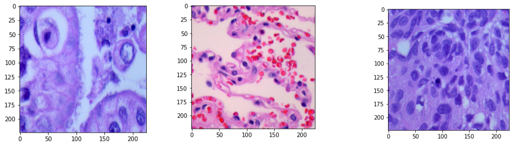
Figure 1: Image samples in the dataset.
The architecture adjustment includes additional dense layers to improve feature representation and classification precision. This adapted network demonstrates significant accuracy improvements by utilizing feature maps through hierarchical representation.
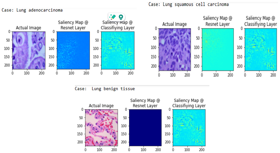
Figure 2: Saliency maps showing features extracted from lung CT images.
Results and Discussion
Training on Google Colab and utilizing TensorFlow, the model achieved a test accuracy of 98.8%. Other metrics such as precision, recall, and F1-score confirmed this high performance. The model distinguished benign from malignant tissues with outstanding precision and identified different cancer subtypes effectively.
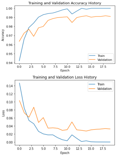
Figure 3: Training and validation curves demonstrating the model's efficiency.
The model's overfitting was minimal, as shown by the congruence of training and validation accuracy and loss curves. A thorough evaluation showed no overfitting tendencies—validation results closely matched training metrics, demonstrating strong generalization capabilities.
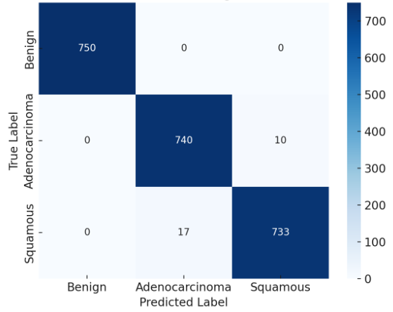
Figure 4: Confusion matrix of the lung cancer prediction model.
Comparative Analysis
The paper provides a comparative study against prior models, demonstrating superior performance. It highlights how ResNet-50, with its deep architecture, can achieve better feature extraction and classification accuracy when adapted appropriately.
| Author |
Model |
Accuracy (%) |
| Garg et al. |
ResNet50 |
96.00 |
| Liang et al. |
Multi-Scale Feature Fusion CNN |
96.00 |
| Shafi et al. |
CNN + SVM |
94.00 |
| tawfeek et al. |
LCRP |
98.50 |
| Kumar et al. |
Fusion Model and ResNet-50 |
90.00 |
| Current Paper |
Adapted ResNet50 |
98.80 |
Conclusion
The paper presents a highly efficient approach for multiclass lung cancer classification through adaptation of the ResNet-50 architecture. The fine-tuned model significantly improves upon existing methods and achieves notable performance in distinguishing between lung cancer subtypes based on CT imaging. Future research directions may include exploring larger datasets and additional cancer classes for enhanced diagnostic utility.