- The paper presents a VGG16-based classifier that differentiates between healthy and diseased muscular tissue with accuracies up to 0.96.
- It integrates photoacoustic and ultrasound imaging, highlighting ultrasound's superior performance in distinguishing disease subtypes.
- The study analyzes disease progression, noting challenges in early-stage DMD detection and the potential for enhanced multi-modal diagnostic approaches.
Automatic Classification of Neuromuscular Diseases in Children Using Photoacoustic Imaging (2201.11630)
Introduction
This paper investigates the automatic classification of neuromuscular diseases (NMDs), specifically Duchenne Muscular Dystrophy (DMD) and Spinal Muscular Atrophy (SMA), in pediatric patients using photoacoustic (PA) imaging combined with ultrasound (US) imaging. The authors propose utilizing deep learning (DL) techniques to classify diseased versus healthy muscular tissue. A VGG16-based classifier was developed to differentiate between healthy states and the disease subtypes, achieving promising accuracy levels that support future applications in medical diagnostics.
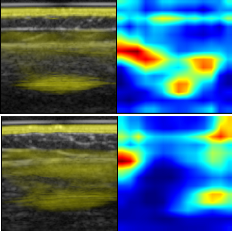
Figure 1: Imaging results for Spinal Muscular Atrophy (SMA) using PA imaging overlaid on ultrasound (US) images.
Methodology
Data Collection and Preprocessing
The dataset includes PA and US images from 10 DMD patients, 10 SMA patients, and 20 healthy controls. Image acquisition was performed using a handheld PA system, capturing 10,683 frames at varying wavelengths (800 nm, 920 nm) or spectrally unmixed signals (SUS). Following preprocessing with vendor-specific software, images were resized to 224×224 px, serving as input for neural networks. Data partitioning into training, validation, and test sets respected a 0.5/0.2/0.3 split to account for class representation.
Experimental Setup
For classification tasks, a VGG16 network, pre-trained on ImageNet, was adapted with four distinct inputs: US, two WL images (800 nm, 920 nm), and SUS images. Only final layers were retrained, employing stochastic gradient descent with momentum. Weighted cross-entropy loss counterbalanced class imbalances. The DL approach was enhanced with random augmentation techniques and image normalization. Class Activation Mapping (Grad-CAM) visually determined network prediction relevance, providing interpretability through heatmaps.
Results
VGG16 networks trained on US images outperformed other modalities, with accuracies reaching 0.96 for DMD class separation and 0.91 for the three-class (healthy, DMD, SMA) classification, as depicted in validation phases. The final test evaluations displayed robust accuracies between 0.86 and 1.00 across tasks (DMD vs. Control, SMA vs. Control, and 3-class scenarios). Notably, US inputs consistently registered higher performance compared to PA-based inputs.
Disease Stage Analysis
The paper reports greater misclassification tendencies in early-stage disease cases, like DMD in younger cohorts and less severe SMA types. An analysis of age versus classification accuracy aligns with DMD’s progression, where older patients present more distinct symptoms, enhancing classification precision.
Discussion
Despite US imaging's superior classification performance, PA imaging introduces functional tissue characterization that complements morphological details observable in ultrasound. Such integrative imaging augments diagnostic resolution, especially in distinguishing NMD subtypes. Furthermore, future endeavors could leverage multi-frame data and combinations of PA and US imaging to amplify diagnostic accuracy.
Further exploratory research involves expansion of the dataset and diversification of DL architectures, such as AlexNet and ResNet, to compare against VGG16's benchmarks. Additionally, refinement of PA imaging parameters and preprocessing techniques could illuminate new pathways for integrating these modalities in broader clinical settings, like inflammatory disease diagnostics.
Conclusion
The research underscores PA imaging's potential for augmenting diagnostic accuracy in pediatric neuromuscular disease classification. Despite challenges, PA's blend of structural and functional insights marks it as a favorable candidate for future NMD diagnostic methodologies. Continued development promises not only a deeper understanding of disease progression but also the potential for tailored treatment strategies.